Disentangling Unusual Catalytic Properties and the Role of the [4Fe-4S] Cluster of Three Endonuclease III from the Extremophile D. radiodurans.
Rollo, F., Borges, P.T., Silveira, C.M., Rosa, M.T.G., Todorovic, S., Moe, E.(2022) Molecules 27
- PubMed: 35807515 Search on PubMedSearch on PubMed Central
- DOI: https://doi.org/10.3390/molecules27134270
- Primary Citation Related Structures:
8A5C, 8A5F, 8A5G - PubMed Abstract:
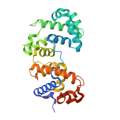
Endonuclease III (EndoIII) is a bifunctional DNA glycosylase with specificity for a broad range of oxidized DNA lesions. The genome of an extremely radiation- and desiccation-resistant bacterium, Deinococcus radiodurans , possesses three genes encoding for EndoIII-like enzymes (DrEndoIII1, DrEndoIII2 and DrEndoIII3), which reveal different types of catalytic activities. DrEndoIII2 acts as the main EndoIII in this organism, while DrEndoIII1 and 3 demonstrate unusual and no EndoIII activity, respectively. In order to understand the role of DrEndoIII1 and DrEndoIII3 in D. radiodurans , we have generated mutants which target non-conserved residues in positions considered essential for classic EndoIII activity. In parallel, we have substituted residues coordinating the iron atoms in the [4Fe-4S] cluster in DrEndoIII2, aiming at elucidating the role of the cluster in these enzymes. Our results demonstrate that the amino acid substitutions in DrEndoIII1 reduce the enzyme activity without altering the overall structure, revealing that the residues found in the wild-type enzyme are essential for its unusual activity. The attempt to generate catalytic activity of DrEndoIII3 by re-designing its catalytic pocket was unsuccessful. A mutation of the iron-coordinating cysteine 199 in DrEndoIII2 appears to compromise the structural integrity and induce the formation of a [3Fe-4S] cluster, but apparently without affecting the activity. Taken together, we provide important structural and mechanistic insights into the three EndoIIIs, which will help us disentangle the open questions related to their presence in D. radiodurans and their particularities.
- Instituto de Tecnologia Química e Biológica António Xavier, Universidade NOVA de Lisboa, Av. da República, 2780-157 Oeiras, Portugal.
Organizational Affiliation: