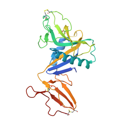
8A59

C-type lectin-like domain (CTLD) and Sushi-like domain of human CD93


Experimental Data Snapshot

  • Method: X-RAY DIFFRACTION
  • Resolution: 1.92 Å
  • R-Value Free: 0.267 
  • R-Value Work: 0.209 
  • R-Value Observed: 0.212 

Starting Model: in silico
View more details

wwPDB Validation   3D Report Full Report


This is version 1.3 of the entry. See complete history


Literature

Dimerization of the C-type lectin-like receptor CD93 promotes its binding to Multimerin-2 in endothelial cells.

Barbera, S.Raucci, L.Tassone, G.Tinti, L.Prischi, F.Santucci, A.Mongiat, M.Tosi, G.M.Galvagni, F.Dimberg, A.Pozzi, C.Orlandini, M.

(2023) Int J Biol Macromol 224: 453-464

  • DOI: https://doi.org/10.1016/j.ijbiomac.2022.10.136
  • Primary Citation of Related Structures:  
    8A59

  • PubMed Abstract: 

    Blocking the signaling activated by the plasma membrane receptor CD93 has recently been demonstrated a useful tool in antiangiogenic treatment and oncotherapy. In the proliferating endothelium, CD93 regulates cell adhesion, migration, and vascular maturation, yet it is unclear how CD93 interacts with the extracellular matrix activating signaling pathways involved in the vascular remodeling. Here for the first time we show that in endothelial cells CD93 is structured as a dimer and that this oligomeric form is physiologically instrumental for the binding of CD93 to its ligand Multimerin-2. Crystallographic X-ray analysis of recombinant CD93 reveals the crucial role played by the C-type lectin-like and sushi-like domains in arranging as an antiparallel dimer to achieve a functional binding state, providing key information for the future design of new drugs able to hamper CD93 function in neovascular pathologies.


  • Organizational Affiliation

    Department of Biotechnology, Chemistry and Pharmacy, University of Siena, Siena 53100, Italy; Department of Immunology, Genetics and Pathology, Science for Life Laboratory, Uppsala University, Rudbeck Laboratory, Uppsala SE-75185, Sweden.


Macromolecules
Find similar proteins by:  (by identity cutoff)  |  3D Structure
Entity ID: 1
MoleculeChains Sequence LengthOrganismDetailsImage
Complement component C1q receptor239Homo sapiensMutation(s): 0 
Gene Names: CD93C1QR1MXRA4
UniProt & NIH Common Fund Data Resources
Find proteins for Q9NPY3 (Homo sapiens)
Explore Q9NPY3 
Go to UniProtKB:  Q9NPY3
PHAROS:  Q9NPY3
GTEx:  ENSG00000125810 
Entity Groups  
Sequence Clusters30% Identity50% Identity70% Identity90% Identity95% Identity100% Identity
UniProt GroupQ9NPY3
Sequence Annotations
Expand
  • Reference Sequence
Small Molecules
Ligands 2 Unique
IDChains Name / Formula / InChI Key2D Diagram3D Interactions
SO4
Query on SO4

Download Ideal Coordinates CCD File 
H [auth A],
I [auth A],
J [auth A]
SULFATE ION
O4 S
QAOWNCQODCNURD-UHFFFAOYSA-L
GOL
Query on GOL

Download Ideal Coordinates CCD File 
B [auth A]
C [auth A]
D [auth A]
E [auth A]
F [auth A]
B [auth A],
C [auth A],
D [auth A],
E [auth A],
F [auth A],
G [auth A]
GLYCEROL
C3 H8 O3
PEDCQBHIVMGVHV-UHFFFAOYSA-N
Experimental Data & Validation

Experimental Data

  • Method: X-RAY DIFFRACTION
  • Resolution: 1.92 Å
  • R-Value Free: 0.267 
  • R-Value Work: 0.209 
  • R-Value Observed: 0.212 
  • Space Group: C 2 2 21
Unit Cell:
Length ( Å )Angle ( ˚ )
a = 63.712α = 90
b = 112.504β = 90
c = 73.262γ = 90
Software Package:
Software NamePurpose
REFMACrefinement
PDB_EXTRACTdata extraction
XDSdata reduction
SCALAdata scaling
MOLREPphasing

Structure Validation

View Full Validation Report


Entry History & Funding Information

Deposition Data


Funding OrganizationLocationGrant Number
Not funded--

Revision History  (Full details and data files)

  • Version 1.0: 2022-11-02
    Type: Initial release
  • Version 1.1: 2022-12-28
    Changes: Database references
  • Version 1.2: 2024-05-01
    Changes: Data collection, Refinement description
  • Version 1.3: 2024-10-16
    Changes: Structure summary