Discovery of Nanomolar-Affinity Pharmacological Chaperones Stabilizing the Oncogenic p53 Mutant Y220C.
Stephenson Clarke, J.R., Douglas, L.R., Duriez, P.J., Balourdas, D.I., Joerger, A.C., Khadiullina, R., Bulatov, E., Baud, M.G.J.(2022) Acs Pharmacol Transl Sci 5: 1169-1180
- PubMed: 36407959 Search on PubMedSearch on PubMed Central
- DOI: https://doi.org/10.1021/acsptsci.2c00164
- Primary Citation Related Structures:
8A31, 8A32 - PubMed Abstract:
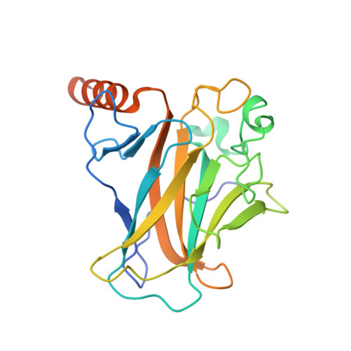
The tumor suppressor protein p53 is inactivated in the majority of human cancers and remains a prime target for developing new drugs to reactivate its tumor suppressing activity for anticancer therapies. The oncogenic p53 mutant Y220C accounts for approximately 125,000 new cancer cases per annum and is one of the most prevalent p53 mutants overall. It harbors a narrow, mutationally induced pocket at the surface of the DNA-binding domain that destabilizes p53, leading to its rapid denaturation and aggregation. Here, we present the structure-guided development of high-affinity small molecules stabilizing p53-Y220C in vitro , along with the synthetic routes developed in the process, in vitro structure-activity relationship data, and confirmation of their binding mode by protein X-ray crystallography. We disclose two new chemical probes displaying sub-micromolar binding affinity in vitro , marking an important milestone since the discovery of the first small-molecule ligand of Y220C in 2008. New chemical probe JC744 displayed a K d = 320 nM, along with potent in vitro protein stabilization. This study, therefore, represents a significant advance toward high-affinity Y220C ligands for clinical evaluation.
- School of Chemistry and Institute for Life Sciences, University of Southampton, Southampton SO17 1BJ, United Kingdom.
Organizational Affiliation: