8A1F

Human PTPRK N-terminal domains MAM-Ig-FN1


Experimental Data Snapshot

  • Method: X-RAY DIFFRACTION
  • Resolution: 3.00 Å
  • R-Value Free: 0.274 
  • R-Value Work: 0.228 
  • R-Value Observed: 0.230 

wwPDB Validation   3D Report Full Report


Ligand Structure Quality Assessment 


This is version 1.1 of the entry. See complete history


Literature

Determinants of receptor tyrosine phosphatase homophilic adhesion: Structural comparison of PTPRK and PTPRM extracellular domains.

Hay, I.M.Shamin, M.Caroe, E.R.Mohammed, A.S.A.Svergun, D.I.Jeffries, C.M.Graham, S.C.Sharpe, H.J.Deane, J.E.

(2023) J Biol Chem 299: 102750-102750

  • DOI: https://doi.org/10.1016/j.jbc.2022.102750
  • Primary Citation of Related Structures:  
    8A16, 8A17, 8A1F

  • PubMed Abstract: 

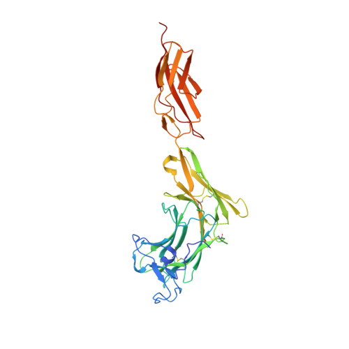
    Type IIB receptor protein tyrosine phosphatases are cell surface transmembrane proteins that engage in cell adhesion via their extracellular domains (ECDs) and cell signaling via their cytoplasmic phosphatase domains. The ECDs of type IIB receptor protein tyrosine phosphatases form stable, homophilic, and trans interactions between adjacent cell membranes. Previous work has demonstrated how one family member, PTPRM, forms head-to-tail homodimers. However, as the interface was composed of residues conserved across the family, the determinants of homophilic specificity remain unknown. Here, we have solved the X-ray crystal structure of the membrane-distal N-terminal domains of PTPRK that form a head-to-tail dimer consistent with intermembrane adhesion. Comparison with the PTPRM structure demonstrates interdomain conformational differences that may define homophilic specificity. Using small-angle X-ray scattering, we determined the solution structures of the full-length ECDs of PTPRM and PTPRK, identifying that both are rigid extended molecules that differ in their overall long-range conformation. Furthermore, we identified one residue, W351, within the interaction interface that differs between PTPRM and PTPRK and showed that mutation to glycine, the equivalent residue in PTPRM, abolishes PTPRK dimer formation in vitro. This comparison of two members of the receptor tyrosine phosphatase family suggests that homophilic specificity is driven by a combination of shape complementarity and specific but limited sequence differences.


  • Organizational Affiliation

    Cambridge Institute for Medical Research, University of Cambridge, Cambridge, United Kingdom; Signalling Programme, Babraham Institute, Babraham Research Campus, Cambridge, United Kingdom.


Macromolecules
Find similar proteins by:  (by identity cutoff)  |  3D Structure
Entity ID: 1
MoleculeChains Sequence LengthOrganismDetailsImage
Receptor-type tyrosine-protein phosphatase kappa
A, B
370Homo sapiensMutation(s): 0 
Gene Names: PTPRKPTPK
EC: 3.1.3.48
UniProt & NIH Common Fund Data Resources
Find proteins for Q15262 (Homo sapiens)
Explore Q15262 
Go to UniProtKB:  Q15262
PHAROS:  Q15262
GTEx:  ENSG00000152894 
Entity Groups  
Sequence Clusters30% Identity50% Identity70% Identity90% Identity95% Identity100% Identity
UniProt GroupQ15262
Sequence Annotations
Expand
  • Reference Sequence
Oligosaccharides

Help

Entity ID: 2
MoleculeChains Length2D Diagram Glycosylation3D Interactions
2-acetamido-2-deoxy-beta-D-glucopyranose-(1-4)-[alpha-L-fucopyranose-(1-6)]2-acetamido-2-deoxy-beta-D-glucopyranose
C
3N-Glycosylation
Glycosylation Resources
GlyTouCan:  G21290RB
GlyCosmos:  G21290RB
GlyGen:  G21290RB
Entity ID: 3
MoleculeChains Length2D Diagram Glycosylation3D Interactions
alpha-D-mannopyranose-(1-3)-beta-D-mannopyranose-(1-4)-2-acetamido-2-deoxy-beta-D-glucopyranose-(1-4)-2-acetamido-2-deoxy-beta-D-glucopyranose
D
4N-Glycosylation
Glycosylation Resources
GlyTouCan:  G81315DD
GlyCosmos:  G81315DD
GlyGen:  G81315DD
Entity ID: 4
MoleculeChains Length2D Diagram Glycosylation3D Interactions
2-acetamido-2-deoxy-beta-D-glucopyranose-(1-4)-2-acetamido-2-deoxy-beta-D-glucopyranose
E
2N-Glycosylation
Glycosylation Resources
GlyTouCan:  G42666HT
GlyCosmos:  G42666HT
GlyGen:  G42666HT
Entity ID: 5
MoleculeChains Length2D Diagram Glycosylation3D Interactions
beta-D-mannopyranose-(1-4)-2-acetamido-2-deoxy-beta-D-glucopyranose-(1-4)-2-acetamido-2-deoxy-beta-D-glucopyranose
F
3N-Glycosylation
Glycosylation Resources
GlyTouCan:  G15407YE
GlyCosmos:  G15407YE
GlyGen:  G15407YE
Experimental Data & Validation

Experimental Data

  • Method: X-RAY DIFFRACTION
  • Resolution: 3.00 Å
  • R-Value Free: 0.274 
  • R-Value Work: 0.228 
  • R-Value Observed: 0.230 
  • Space Group: P 21 21 21
Unit Cell:
Length ( Å )Angle ( ˚ )
a = 87.79α = 90
b = 93.862β = 90
c = 181.737γ = 90
Software Package:
Software NamePurpose
PHENIXrefinement
PHASERphasing
DIALSdata scaling
Cootmodel building
DIALSdata reduction
DIALSdata scaling

Structure Validation

View Full Validation Report


Ligand Structure Quality Assessment 


Entry History & Funding Information

Deposition Data


Funding OrganizationLocationGrant Number
Wellcome TrustUnited Kingdom109407/Z/15/Z
Wellcome TrustUnited Kingdom219447/Z/19/Z

Revision History  (Full details and data files)

  • Version 1.0: 2023-03-15
    Type: Initial release
  • Version 1.1: 2024-02-07
    Changes: Data collection, Refinement description