8A0F

Crystal structure of human deoxyhypusine synthase variant K329A in complex with NAD and SPD


Experimental Data Snapshot

  • Method: X-RAY DIFFRACTION
  • Resolution: 1.64 Å
  • R-Value Free: 0.178 
  • R-Value Work: 0.156 
  • R-Value Observed: 0.156 

wwPDB Validation   3D Report Full Report


Ligand Structure Quality Assessment 


This is version 1.1 of the entry. See complete history


Literature

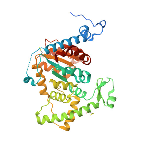
Cryo-EM structure of human eIF5A-DHS complex reveals the molecular basis of hypusination-associated neurodegenerative disorders.

Wator, E.Wilk, P.Biela, A.Rawski, M.Zak, K.M.Steinchen, W.Bange, G.Glatt, S.Grudnik, P.

(2023) Nat Commun 14: 1698-1698

  • DOI: https://doi.org/10.1038/s41467-023-37305-2
  • Primary Citation of Related Structures:  
    7A6S, 7A6T, 8A0E, 8A0F, 8A0G

  • PubMed Abstract: 

    Hypusination is a unique post-translational modification of the eukaryotic translation factor 5A (eIF5A) that is essential for overcoming ribosome stalling at polyproline sequence stretches. The initial step of hypusination, the formation of deoxyhypusine, is catalyzed by deoxyhypusine synthase (DHS), however, the molecular details of the DHS-mediated reaction remained elusive. Recently, patient-derived variants of DHS and eIF5A have been linked to rare neurodevelopmental disorders. Here, we present the cryo-EM structure of the human eIF5A-DHS complex at 2.8 Å resolution and a crystal structure of DHS trapped in the key reaction transition state. Furthermore, we show that disease-associated DHS variants influence the complex formation and hypusination efficiency. Hence, our work dissects the molecular details of the deoxyhypusine synthesis reaction and reveals how clinically-relevant mutations affect this crucial cellular process.


  • Organizational Affiliation

    Małopolska Centre of Biotechnology, Jagiellonian University, Kraków, Poland.


Macromolecules
Find similar proteins by:  (by identity cutoff)  |  3D Structure
Entity ID: 1
MoleculeChains Sequence LengthOrganismDetailsImage
Deoxyhypusine synthase
A, B
371Homo sapiensMutation(s): 1 
Gene Names: DHPSDS
EC: 2.5.1.46
UniProt & NIH Common Fund Data Resources
Find proteins for P49366 (Homo sapiens)
Explore P49366 
Go to UniProtKB:  P49366
PHAROS:  P49366
GTEx:  ENSG00000095059 
Entity Groups  
Sequence Clusters30% Identity50% Identity70% Identity90% Identity95% Identity100% Identity
UniProt GroupP49366
Sequence Annotations
Expand
  • Reference Sequence
Small Molecules
Modified Residues  1 Unique
IDChains TypeFormula2D DiagramParent
CSS
Query on CSS
A, B
L-PEPTIDE LINKINGC3 H7 N O2 S2CYS
Experimental Data & Validation

Experimental Data

  • Method: X-RAY DIFFRACTION
  • Resolution: 1.64 Å
  • R-Value Free: 0.178 
  • R-Value Work: 0.156 
  • R-Value Observed: 0.156 
  • Space Group: P 32 2 1
Unit Cell:
Length ( Å )Angle ( ˚ )
a = 105.241α = 90
b = 105.241β = 90
c = 160.22γ = 120
Software Package:
Software NamePurpose
PHENIXrefinement
XSCALEdata scaling
PDB_EXTRACTdata extraction
PHASERphasing
XDSdata reduction

Structure Validation

View Full Validation Report


Ligand Structure Quality Assessment 


Entry History & Funding Information

Deposition Data


Funding OrganizationLocationGrant Number
Polish National Science CentrePolandUMO-2019/33/B/NZ1/01839
Foundation for Polish SciencePolandTEAM TECH CORE FACILITY/2017-4/6

Revision History  (Full details and data files)

  • Version 1.0: 2023-04-05
    Type: Initial release
  • Version 1.1: 2024-02-07
    Changes: Data collection, Refinement description