8A00

Infectious mouse-adapted ME7 scrapie prion fibril purified from terminally-infected mouse brains


Experimental Data Snapshot

  • Method: ELECTRON MICROSCOPY
  • Resolution: 2.60 Å
  • Aggregation State: FILAMENT 
  • Reconstruction Method: HELICAL 

wwPDB Validation   3D Report Full Report


This is version 1.2 of the entry. See complete history


Literature

A structural basis for prion strain diversity.

Manka, S.W.Wenborn, A.Betts, J.Joiner, S.Saibil, H.R.Collinge, J.Wadsworth, J.D.F.

(2023) Nat Chem Biol 19: 607-613

  • DOI: https://doi.org/10.1038/s41589-022-01229-7
  • Primary Citation of Related Structures:  
    8A00

  • PubMed Abstract: 

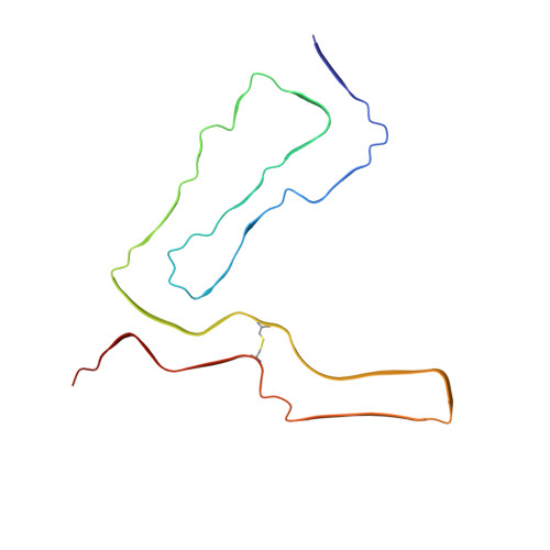
    Recent cryogenic electron microscopy (cryo-EM) studies of infectious, ex vivo, prion fibrils from hamster 263K and mouse RML prion strains revealed a similar, parallel in-register intermolecular β-sheet (PIRIBS) amyloid architecture. Rungs of the fibrils are composed of individual prion protein (PrP) monomers that fold to create distinct N-terminal and C-terminal lobes. However, disparity in the hamster/mouse PrP sequence precludes understanding of how divergent prion strains emerge from an identical PrP substrate. In this study, we determined the near-atomic resolution cryo-EM structure of infectious, ex vivo mouse prion fibrils from the ME7 prion strain and compared this with the RML fibril structure. This structural comparison of two biologically distinct mouse-adapted prion strains suggests defined folding subdomains of PrP rungs and the way in which they are interrelated, providing a structural definition of intra-species prion strain-specific conformations.


  • Organizational Affiliation

    MRC Prion Unit at UCL, Institute of Prion Diseases, University College London, London, UK.


Macromolecules
Find similar proteins by:  (by identity cutoff)  |  3D Structure
Entity ID: 1
MoleculeChains Sequence LengthOrganismDetailsImage
Major prion proteinA,
B [auth C],
C [auth B]
136Mus musculusMutation(s): 0 
UniProt
Find proteins for P04925 (Mus musculus)
Explore P04925 
Go to UniProtKB:  P04925
Entity Groups  
Sequence Clusters30% Identity50% Identity70% Identity90% Identity95% Identity100% Identity
UniProt GroupP04925
Sequence Annotations
Expand
  • Reference Sequence
Experimental Data & Validation

Experimental Data

  • Method: ELECTRON MICROSCOPY
  • Resolution: 2.60 Å
  • Aggregation State: FILAMENT 
  • Reconstruction Method: HELICAL 
EM Software:
TaskSoftware PackageVersion
RECONSTRUCTIONRELION4.0-beta
MODEL REFINEMENTPHENIX

Structure Validation

View Full Validation Report


Entry History & Funding Information

Deposition Data


Funding OrganizationLocationGrant Number
Medical Research Council (MRC, United Kingdom)United KingdomMC_U12316055
Medical Research Council (MRC, United Kingdom)United KingdomMC_UU_00024/5
Wellcome TrustUnited Kingdom(202679/Z/16/Z
Wellcome TrustUnited Kingdom206166/Z/17/Z
Wellcome TrustUnited Kingdom106249/Z/14/Z

Revision History  (Full details and data files)

  • Version 1.0: 2023-01-18
    Type: Initial release
  • Version 1.1: 2023-02-08
    Changes: Database references
  • Version 1.2: 2023-05-10
    Changes: Database references