7ZU6

Polyoxidovanadate interaction with proteins: crystal structure of lysozyme bound to tetra-vanadate ion (structure 1)


Experimental Data Snapshot

  • Method: X-RAY DIFFRACTION
  • Resolution: 1.18 Å
  • R-Value Free: 0.196 
  • R-Value Work: 0.171 

wwPDB Validation   3D Report Full Report


This is version 1.3 of the entry. See complete history


Literature

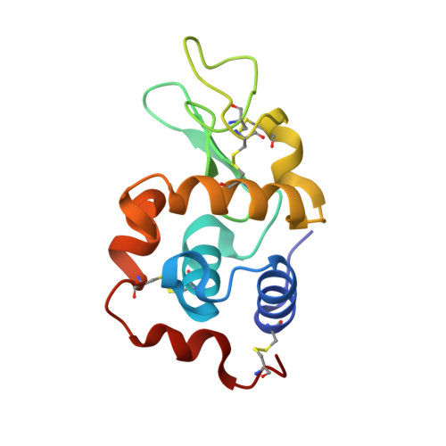
Stabilization and Binding of [V 4 O 12 ] 4- and Unprecedented [V 20 O 54 (NO 3 )] n- to Lysozyme upon Loss of Ligands and Oxidation of the Potential Drug V IV O(acetylacetonato) 2.

Ferraro, G.Tito, G.Sciortino, G.Garribba, E.Merlino, A.

(2023) Angew Chem Int Ed Engl 62: e202310655-e202310655

  • DOI: https://doi.org/10.1002/anie.202310655
  • Primary Citation of Related Structures:  
    7ZU6, 8PTE

  • PubMed Abstract: 

    High-resolution crystal structures of lysozyme in the presence of the potential drug V IV O(acetylacetonato) 2 under two different experimental conditions have been solved. The crystallographic study reveals the loss of the ligands, the oxidation of V IV to V V and the subsequent formation of adducts of the protein with two different polyoxidovanadates: [V 4 O 12 ] 4- , which interacts with lysozyme non-covalently, and the unprecedented [V 20 O 54 (NO 3 )] n- , which is covalenty bound to the side chain of an aspartate residue of symmetry related molecules.


  • Organizational Affiliation

    Department of Chemical Sciences, University of Naples Federico II, Complesso Universitario di Monte Sant'Angelo, Via Cintia, I-80126, Napoli, Italy.


Macromolecules
Find similar proteins by:  (by identity cutoff)  |  3D Structure
Entity ID: 1
MoleculeChains Sequence LengthOrganismDetailsImage
LysozymeA [auth AAA]129Gallus gallusMutation(s): 0 
EC: 3.2.1.17
UniProt
Find proteins for P00698 (Gallus gallus)
Explore P00698 
Go to UniProtKB:  P00698
Entity Groups  
Sequence Clusters30% Identity50% Identity70% Identity90% Identity95% Identity100% Identity
UniProt GroupP00698
Sequence Annotations
Expand
  • Reference Sequence
Experimental Data & Validation

Experimental Data

  • Method: X-RAY DIFFRACTION
  • Resolution: 1.18 Å
  • R-Value Free: 0.196 
  • R-Value Work: 0.171 
  • Space Group: P 43 21 2
Unit Cell:
Length ( Å )Angle ( ˚ )
a = 77.05α = 90
b = 77.05β = 90
c = 37.84γ = 90
Software Package:
Software NamePurpose
REFMACrefinement
autoPROCdata reduction
autoPROCdata scaling
PHASERphasing

Structure Validation

View Full Validation Report


Entry History & Funding Information

Deposition Data


Funding OrganizationLocationGrant Number
Not funded--

Revision History  (Full details and data files)

  • Version 1.0: 2023-05-24
    Type: Initial release
  • Version 1.1: 2023-11-01
    Changes: Data collection, Database references
  • Version 1.2: 2023-12-13
    Changes: Database references
  • Version 1.3: 2024-02-07
    Changes: Refinement description