7ZSQ | pdb_00007zsq

human purine nucleoside phosphorylase in complex with JS-555


Experimental Data Snapshot

  • Method: X-RAY DIFFRACTION
  • Resolution: 1.77 Å
  • R-Value Free: 
    0.214 (Depositor), 0.224 (DCC) 
  • R-Value Work: 
    0.182 (Depositor), 0.192 (DCC) 
  • R-Value Observed: 
    0.182 (Depositor) 

Starting Model: experimental
View more details

wwPDB Validation 3D Report Full Report

Validation slider image for 7ZSQ

Ligand Structure Quality Assessment 


This is version 1.3 of the entry. See complete history

Literature

Design, Synthesis, Biological Evaluation, and Crystallographic Study of Novel Purine Nucleoside Phosphorylase Inhibitors.

Skacel, J.Djukic, S.Baszczynski, O.Kalcic, F.Bilek, T.Chalupsky, K.Kozak, J.Dvorakova, A.Tloust'ova, E.Kral'ova, Z.Smidkova, M.Voldrich, J.Rumlova, M.Pachl, P.Brynda, J.Vuckova, T.Fabry, M.Snasel, J.Pichova, I.Rezacova, P.Mertlikova-Kaiserova, H.Janeba, Z.

(2023) J Med Chem 66: 6652-6681

  • DOI: https://doi.org/10.1021/acs.jmedchem.2c02097
  • Primary Citation Related Structures: 
    7ZSL, 7ZSM, 7ZSN, 7ZSO, 7ZSP, 7ZSQ, 7ZSR, 8C25

  • PubMed Abstract: 

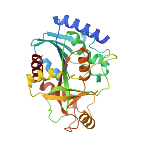
    Purine nucleoside phosphorylase (PNP) is a well-known molecular target with potential therapeutic applications in the treatment of T-cell malignancies and/or bacterial/parasitic infections. Here, we report the design, development of synthetic methodology, and biological evaluation of a series of 30 novel PNP inhibitors based on acyclic nucleoside phosphonates bearing a 9-deazahypoxanthine nucleobase. The strongest inhibitors exhibited IC 50 values as low as 19 nM (human PNP) and 4 nM ( Mycobacterium tuberculosis ( Mt ) PNP) and highly selective cytotoxicity toward various T-lymphoblastic cell lines with CC 50 values as low as 9 nM. No cytotoxic effect was observed on other cancer cell lines (HeLa S3, HL60, HepG2) or primary PBMCs for up to 10 μM. We report the first example of the PNP inhibitor exhibiting over 60-fold selectivity for the pathogenic enzyme ( Mt PNP) over hPNP. The results are supported by a crystallographic study of eight enzyme-inhibitor complexes and by ADMET profiling in vitro and in vivo .


  • Organizational Affiliation
    • Institute of Organic Chemistry and Biochemistry, The Czech Academy of Sciences, Flemingovo nám. 2, Prague 16610, Czech Republic.

Macromolecule Content 

  • Total Structure Weight: 86.77 kDa 
  • Atom Count: 6,260 
  • Modeled Residue Count: 791 
  • Deposited Residue Count: 807 
  • Unique protein chains: 1

Macromolecules

Find similar proteins by:|  3D Structure
Entity ID: 1
MoleculeChains  Sequence LengthOrganismDetailsImage
Purine nucleoside phosphorylase
A, B, C
269Mycobacterium tuberculosisMutation(s): 0 
Gene Names: deoDE5M05_03615E5M23_14660E5M52_18960E5M78_19105ERS013440_01955ERS027646_00621ERS027659_03654SAMEA2683035_02840
EC: 2.4.2.1
UniProt
Find proteins for P9WP01 (Mycobacterium tuberculosis (strain ATCC 25618 / H37Rv))
Explore P9WP01 
Go to UniProtKB:  P9WP01
Entity Groups
Sequence Clusters30% Identity50% Identity70% Identity90% Identity95% Identity100% Identity
UniProt GroupP9WP01
Sequence Annotations
Expand
Reference Sequence

Small Molecules

Ligands 6 Unique
IDChains Name / Formula / InChI Key2D Diagram3D Interactions
TEW

Query on TEW


Download:Ideal Coordinates CCD File
E [auth A]6-tungstotellurate(VI)
O24 Te W6
JDLYBIMRCSJRQJ-UHFFFAOYSA-M
P4K

Query on P4K


Download:Ideal Coordinates CCD File
H [auth B]polyethylene glycol
C30 H62 O15
WWPGFZAAWXFBTF-UHFFFAOYSA-N
JTO
(Subject of Investigation/LOI)

Query on JTO


Download:Ideal Coordinates CCD File
D [auth A],
G [auth B],
J [auth C]
[2-[(4-oxidanylidene-3,5-dihydropyrrolo[3,2-d]pyrimidin-7-yl)sulfanyl]phenoxy]methylphosphonic acid
C13 H12 N3 O5 P S
VOAJLIGXNWIOPN-UHFFFAOYSA-N
PEG

Query on PEG


Download:Ideal Coordinates CCD File
K [auth C]DI(HYDROXYETHYL)ETHER
C4 H10 O3
MTHSVFCYNBDYFN-UHFFFAOYSA-N
GOL

Query on GOL


Download:Ideal Coordinates CCD File
F [auth A]GLYCEROL
C3 H8 O3
PEDCQBHIVMGVHV-UHFFFAOYSA-N
EDO

Query on EDO


Download:Ideal Coordinates CCD File
I [auth B]1,2-ETHANEDIOL
C2 H6 O2
LYCAIKOWRPUZTN-UHFFFAOYSA-N

Experimental Data & Validation

Experimental Data

  • Method: X-RAY DIFFRACTION
  • Resolution: 1.77 Å
  • R-Value Free:  0.214 (Depositor), 0.224 (DCC) 
  • R-Value Work:  0.182 (Depositor), 0.192 (DCC) 
  • R-Value Observed: 0.182 (Depositor) 
Space Group: P 21 21 21
Unit Cell:
Length ( Å )Angle ( ˚ )
a = 57.07α = 90
b = 90.93β = 90
c = 143.07γ = 90
Software Package:
Software NamePurpose
XSCALEdata scaling
REFMACrefinement
PDB_EXTRACTdata extraction
XDSdata reduction

Structure Validation

View Full Validation Report


Ligand Structure Quality Assessment 


Entry History 

& Funding Information

Deposition Data


Funding OrganizationLocationGrant Number
European Regional Development FundEuropean UnionCZ.02.1.01/0.0/0.0/16_019/0000729

Revision History  (Full details and data files)

  • Version 1.0: 2023-05-17
    Type: Initial release
  • Version 1.1: 2023-05-24
    Changes: Database references
  • Version 1.2: 2023-06-07
    Changes: Database references
  • Version 1.3: 2024-02-07
    Changes: Data collection, Refinement description