7ZRP

2.65 Angstrom crystal structure of Ca/CaM:CaMKIIdelta peptide complex


Experimental Data Snapshot

  • Method: X-RAY DIFFRACTION
  • Resolution: 2.65 Å
  • R-Value Free: 0.272 
  • R-Value Work: 0.207 

wwPDB Validation   3D Report Full Report


This is version 1.1 of the entry. See complete history


Literature

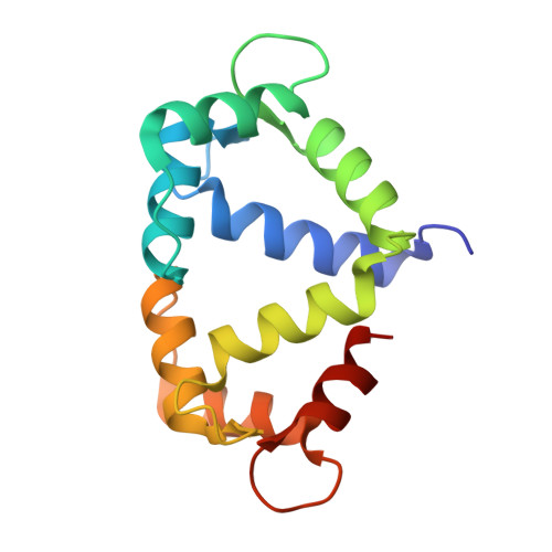
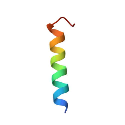
Calmodulin variant E140G associated with long QT syndrome impairs CaMKII delta autophosphorylation and L-type calcium channel inactivation.

Prakash, O.Gupta, N.Milburn, A.McCormick, L.Deugi, V.Fisch, P.Wyles, J.Thomas, N.L.Antonyuk, S.Dart, C.Helassa, N.

(2022) J Biol Chem 299: 102777-102777

  • DOI: https://doi.org/10.1016/j.jbc.2022.102777
  • Primary Citation of Related Structures:  
    7ZRP, 7ZRQ

  • PubMed Abstract: 
  • Long QT syndrome (LQTS) is a human inherited heart condition that can cause life-threatening arrhythmia including sudden cardiac death. Mutations in the ubiquitous Ca 2+ -sensing protein calmodulin (CaM) are associated with LQTS, but the molecular mechanism by which these mutations lead to irregular heartbeats is not fully understood ...

    Long QT syndrome (LQTS) is a human inherited heart condition that can cause life-threatening arrhythmia including sudden cardiac death. Mutations in the ubiquitous Ca 2+ -sensing protein calmodulin (CaM) are associated with LQTS, but the molecular mechanism by which these mutations lead to irregular heartbeats is not fully understood. Here, we use a multidisciplinary approach including protein biophysics, structural biology, confocal imaging, and patch-clamp electrophysiology to determine the effect of the disease-associated CaM mutation E140G on CaM structure and function. We present novel data showing that mutant-regulated CaMKIIδ kinase activity is impaired with a significant reduction in enzyme autophosphorylation rate. We report the first high-resolution crystal structure of a LQTS-associated CaM variant in complex with the CaMKIIδ peptide, which shows significant structural differences, compared to the WT complex. Furthermore, we demonstrate that the E140G mutation significantly disrupted Ca v 1.2 Ca 2+ /CaM-dependent inactivation, while cardiac ryanodine receptor (RyR2) activity remained unaffected. In addition, we show that the LQTS-associated mutation alters CaM's Ca 2+ -binding characteristics, secondary structure content, and interaction with key partners involved in excitation-contraction coupling (CaMKIIδ, Ca v 1.2, RyR2). In conclusion, LQTS-associated CaM mutation E140G severely impacts the structure-function relationship of CaM and its regulation of CaMKIIδ and Ca v 1.2. This provides a crucial insight into the molecular factors contributing to CaM-mediated arrhythmias with a central role for CaMKIIδ.


    Organizational Affiliation

    Liverpool Centre for Cardiovascular Science, Department of Cardiovascular and Metabolic Medicine, Institute of Life Course and Medical Sciences, Faculty of Health and Life Sciences, University of Liverpool, Liverpool, United Kingdom. Electronic address: nhelassa@liverpool.ac.uk.


Macromolecules
Find similar proteins by:  (by identity cutoff)  |  3D Structure
Entity ID: 1
MoleculeChainsSequence LengthOrganismDetailsImage
Calmodulin-1
A, C
148Homo sapiensMutation(s): 0 
Gene Names: CALM1CALMCAMCAM1
UniProt & NIH Common Fund Data Resources
Find proteins for P0DP23 (Homo sapiens)
Explore P0DP23 
Go to UniProtKB:  P0DP23
PHAROS:  P0DP23
Entity Groups  
Sequence Clusters30% Identity50% Identity70% Identity90% Identity95% Identity100% Identity
UniProt GroupP0DP23
Protein Feature View
Expand
  • Reference Sequence

Find similar proteins by:  Sequence   |   3D Structure  

Entity ID: 2
MoleculeChainsSequence LengthOrganismDetailsImage
Calcium/calmodulin-dependent protein kinase type II subunit delta
B, D
22Homo sapiensMutation(s): 0 
Gene Names: CAMK2DCAMKD
EC: 2.7.11.17
UniProt & NIH Common Fund Data Resources
Find proteins for Q13557 (Homo sapiens)
Explore Q13557 
Go to UniProtKB:  Q13557
PHAROS:  Q13557
Entity Groups  
Sequence Clusters30% Identity50% Identity70% Identity90% Identity95% Identity100% Identity
UniProt GroupQ13557
Protein Feature View
Expand
  • Reference Sequence
Small Molecules
Ligands 4 Unique
IDChainsName / Formula / InChI Key2D Diagram3D Interactions
PG4
Query on PG4

Download Ideal Coordinates CCD File 
I [auth A],
J [auth A]
TETRAETHYLENE GLYCOL
C8 H18 O5
UWHCKJMYHZGTIT-UHFFFAOYSA-N
 Ligand Interaction
IMD
Query on IMD

Download Ideal Coordinates CCD File 
K [auth A],
U [auth C]
IMIDAZOLE
C3 H5 N2
RAXXELZNTBOGNW-UHFFFAOYSA-O
 Ligand Interaction
ZN
Query on ZN

Download Ideal Coordinates CCD File 
L [auth A]
M [auth A]
N [auth A]
O [auth A]
P [auth A]
L [auth A],
M [auth A],
N [auth A],
O [auth A],
P [auth A],
V [auth C],
W [auth C],
X [auth C],
Y [auth C]
ZINC ION
Zn
PTFCDOFLOPIGGS-UHFFFAOYSA-N
 Ligand Interaction
CA (Subject of Investigation/LOI)
Query on CA

Download Ideal Coordinates CCD File 
E [auth A]
F [auth A]
G [auth A]
H [auth A]
Q [auth C]
E [auth A],
F [auth A],
G [auth A],
H [auth A],
Q [auth C],
R [auth C],
S [auth C],
T [auth C]
CALCIUM ION
Ca
BHPQYMZQTOCNFJ-UHFFFAOYSA-N
 Ligand Interaction
Experimental Data & Validation

Experimental Data

  • Method: X-RAY DIFFRACTION
  • Resolution: 2.65 Å
  • R-Value Free: 0.272 
  • R-Value Work: 0.207 
  • Space Group: P 21 21 21
Unit Cell:
Length ( Å )Angle ( ˚ )
a = 64.283α = 90
b = 72.59β = 90
c = 78.156γ = 90
Software Package:
Software NamePurpose
DIALSdata reduction
Aimlessdata scaling
MOLREPphasing
REFMACrefinement

Structure Validation

View Full Validation Report


Entry History & Funding Information

Deposition Data


Funding OrganizationLocationGrant Number
British Heart FoundationUnited KingdomFS/17/56/32925
Wellcome TrustUnited Kingdom102172/B/13/Z

Revision History  (Full details and data files)

  • Version 1.0: 2022-12-28
    Type: Initial release
  • Version 1.1: 2023-01-11
    Changes: Database references