7ZLJ

Cryo-EM structure of C-mannosyltransferase CeDPY19, in ternary complex with Dol25-P-C-Man and acceptor peptide, bound to CMT2-Fab and anti-Fab nanobody


Experimental Data Snapshot

  • Method: ELECTRON MICROSCOPY
  • Resolution: 3.63 Å
  • Aggregation State: 3D ARRAY 
  • Reconstruction Method: SINGLE PARTICLE 

wwPDB Validation   3D Report Full Report


This is version 1.2 of the entry. See complete history


Literature

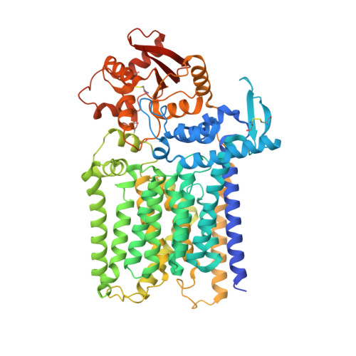
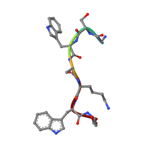
Structure, sequon recognition and mechanism of tryptophan C-mannosyltransferase.

Bloch, J.S.John, A.Mao, R.Mukherjee, S.Boilevin, J.Irobalieva, R.N.Darbre, T.Scott, N.E.Reymond, J.L.Kossiakoff, A.A.Goddard-Borger, E.D.Locher, K.P.

(2023) Nat Chem Biol 19: 575-584

  • DOI: https://doi.org/10.1038/s41589-022-01219-9
  • Primary Citation of Related Structures:  
    7ZLG, 7ZLH, 7ZLI, 7ZLJ

  • PubMed Abstract: 

    C-linked glycosylation is essential for the trafficking, folding and function of secretory and transmembrane proteins involved in cellular communication processes. The tryptophan C-mannosyltransferase (CMT) enzymes that install the modification attach a mannose to the first tryptophan of WxxW/C sequons in nascent polypeptide chains by an unknown mechanism. Here, we report cryogenic-electron microscopy structures of Caenorhabditis elegans CMT in four key states: apo, acceptor peptide-bound, donor-substrate analog-bound and as a trapped ternary complex with both peptide and a donor-substrate mimic bound. The structures indicate how the C-mannosylation sequon is recognized by this CMT and its paralogs, and how sequon binding triggers conformational activation of the donor substrate: a process relevant to all glycosyltransferase C superfamily enzymes. Our structural data further indicate that the CMTs adopt an unprecedented electrophilic aromatic substitution mechanism to enable the C-glycosylation of proteins. These results afford opportunities for understanding human disease and therapeutic targeting of specific CMT paralogs.


  • Organizational Affiliation

    Institute of Molecular Biology and Biophysics, ETH Zürich, Zürich, Switzerland.


Macromolecules
Find similar proteins by:  (by identity cutoff)  |  3D Structure
Entity ID: 1
MoleculeChains Sequence LengthOrganismDetailsImage
CMT2-Fab heavy chainA [auth H]236synthetic constructMutation(s): 0 
Entity Groups  
Sequence Clusters30% Identity50% Identity70% Identity90% Identity95% Identity100% Identity
Sequence Annotations
Expand
  • Reference Sequence
Find similar proteins by:  (by identity cutoff)  |  3D Structure
Entity ID: 2
MoleculeChains Sequence LengthOrganismDetailsImage
Anti-Fab nanobodyB [auth K]123synthetic constructMutation(s): 0 
Entity Groups  
Sequence Clusters30% Identity50% Identity70% Identity90% Identity95% Identity100% Identity
Sequence Annotations
Expand
  • Reference Sequence
Find similar proteins by:  (by identity cutoff)  |  3D Structure
Entity ID: 3
MoleculeChains Sequence LengthOrganismDetailsImage
CMT2-Fab light chainC [auth L]215synthetic constructMutation(s): 0 
Membrane Entity: Yes 
UniProt
Find proteins for Q7Z3Y4 (Homo sapiens)
Explore Q7Z3Y4 
Go to UniProtKB:  Q7Z3Y4
Entity Groups  
Sequence Clusters30% Identity50% Identity70% Identity90% Identity95% Identity100% Identity
UniProt GroupQ7Z3Y4
Sequence Annotations
Expand
  • Reference Sequence
Find similar proteins by:  (by identity cutoff)  |  3D Structure
Entity ID: 4
MoleculeChains Sequence LengthOrganismDetailsImage
C-mannosyltransferase dpy-19D [auth A]707Caenorhabditis elegansMutation(s): 0 
Gene Names: dpy-19F22B7.10
EC: 2.4.1
Membrane Entity: Yes 
UniProt
Find proteins for P34413 (Caenorhabditis elegans)
Explore P34413 
Go to UniProtKB:  P34413
Entity Groups  
Sequence Clusters30% Identity50% Identity70% Identity90% Identity95% Identity100% Identity
UniProt GroupP34413
Sequence Annotations
Expand
  • Reference Sequence

Find similar proteins by:  Sequence   |   3D Structure  

Entity ID: 5
MoleculeChains Sequence LengthOrganismDetailsImage
synthetic octapeptide WEHI 1886493E [auth P]7synthetic constructMutation(s): 0 
Sequence Annotations
Expand
  • Reference Sequence
Small Molecules
Ligands 1 Unique
IDChains Name / Formula / InChI Key2D Diagram3D Interactions
IZU (Subject of Investigation/LOI)
Query on IZU

Download Ideal Coordinates CCD File 
F [auth A][(2~{R},3~{S},4~{S},5~{S},6~{R})-6-(hydroxymethyl)-3,4,5-tris(oxidanyl)oxan-2-yl]methyl-[(3~{S},6~{E},10~{Z},14~{E})-3,7,11,15,19-pentamethylicosa-6,10,14,18-tetraenoxy]phosphinic acid
C32 H57 O8 P
MNRXJRFNBXECBJ-QWFISOLMSA-N
Experimental Data & Validation

Experimental Data

  • Method: ELECTRON MICROSCOPY
  • Resolution: 3.63 Å
  • Aggregation State: 3D ARRAY 
  • Reconstruction Method: SINGLE PARTICLE 

Structure Validation

View Full Validation Report


Entry History & Funding Information

Deposition Data


Funding OrganizationLocationGrant Number
Swiss National Science FoundationSwitzerland--

Revision History  (Full details and data files)

  • Version 1.0: 2023-01-11
    Type: Initial release
  • Version 1.1: 2023-01-18
    Changes: Database references
  • Version 1.2: 2023-05-10
    Changes: Database references