7ZGY

S-layer Deinoxanthin Binding Complex, C3 symmetry


Experimental Data Snapshot

  • Method: ELECTRON MICROSCOPY
  • Resolution: 2.54 Å
  • Aggregation State: PARTICLE 
  • Reconstruction Method: SINGLE PARTICLE 

wwPDB Validation   3D Report Full Report


This is version 1.0 of the entry. See complete history

Re-refinement Note

A newer entry is available that reflects an alternative modeling of the original data: 8ACQ


Literature

The cryo-EM structure of the S-layer deinoxanthin-binding complex of Deinococcus radiodurans informs properties of its environmental interactions.

Farci, D.Haniewicz, P.de Sanctis, D.Iesu, L.Kereiche, S.Winterhalter, M.Piano, D.

(2022) J Biol Chem 298: 102031-102031

  • DOI: https://doi.org/10.1016/j.jbc.2022.102031
  • Primary Citation of Related Structures:  
    7ZGX, 7ZGY

  • PubMed Abstract: 

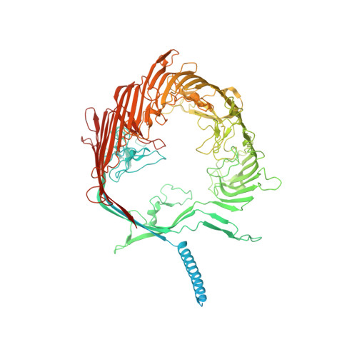
    The radiation-resistant bacterium Deinococcus radiodurans is known as the world's toughest bacterium. The S-layer of D. radiodurans, consisting of several proteins on the surface of the cellular envelope and intimately associated with the outer membrane, has therefore been useful as a model for structural and functional studies. Its main proteinaceous unit, the S-layer deinoxanthin-binding complex (SDBC), is a hetero-oligomeric assembly known to contribute to the resistance against environmental stress and have porin functional features; however, its precise structure is unknown. Here, we resolved the structure of the SDBC at ∼2.5 Å resolution by cryo-EM and assigned the sequence of its main subunit, the protein DR_2577. This structure is characterized by a pore region, a massive β-barrel organization, a stalk region consisting of a trimeric coiled coil, and a collar region at the base of the stalk. We show that each monomer binds three Cu ions and one Fe ion and retains one deinoxanthin molecule and two phosphoglycolipids, all exclusive to D. radiodurans. Finally, electrophysiological characterization of the SDBC shows that it exhibits transport properties with several amino acids. Taken together, these results highlight the SDBC as a robust structure displaying both protection and sieving functions that facilitates exchanges with the environment.


  • Organizational Affiliation

    Department of Plant Physiology, Warsaw University of Life Sciences - SGGW, Warsaw, Poland; Department of Chemistry, Umeå University, Umeå, Sweden. Electronic address: domenica.farci@umu.se.


Macromolecules
Find similar proteins by:  (by identity cutoff)  |  3D Structure
Entity ID: 1
MoleculeChains Sequence LengthOrganismDetailsImage
S-layer protein SlpA
A, B, C
1,167Deinococcus radiodurans R1 = ATCC 13939 = DSM 20539Mutation(s): 0 
Membrane Entity: Yes 
UniProt
Find proteins for Q9RRB6 (Deinococcus radiodurans (strain ATCC 13939 / DSM 20539 / JCM 16871 / CCUG 27074 / LMG 4051 / NBRC 15346 / NCIMB 9279 / VKM B-1422 / R1))
Explore Q9RRB6 
Go to UniProtKB:  Q9RRB6
Entity Groups  
Sequence Clusters30% Identity50% Identity70% Identity90% Identity95% Identity100% Identity
UniProt GroupQ9RRB6
Sequence Annotations
Expand
  • Reference Sequence
Small Molecules
Ligands 5 Unique
IDChains Name / Formula / InChI Key2D Diagram3D Interactions
JPX
Query on JPX

Download Ideal Coordinates CCD File 
F [auth A],
K [auth B],
M [auth B]
[(2~{S})-3-[[(2~{S})-3-[(2~{S},3~{S},4~{S},5~{S},6~{S})-6-(hydroxymethyl)-4,5-bis(oxidanyl)-3-(propanoylamino)oxan-2-yl]oxy-1-oxidanylidene-1-(pentadecylamino)propan-2-yl]oxy-oxidanyl-phosphoryl]oxy-2-octanoyloxy-propyl] decanoate
C48 H91 N2 O15 P
BORXCJRMZBMSCX-GUINJWSRSA-N
JQ6
Query on JQ6

Download Ideal Coordinates CCD File 
E [auth A],
N [auth B],
T [auth C]
[(2~{S})-2-acetyloxy-3-[[(2~{S})-3-[(2~{R},3~{S},4~{S},5~{S},6~{S})-6-(hydroxymethyl)-3-(octadecanoylamino)-4,5-bis(oxidanyl)oxan-2-yl]oxy-1-oxidanylidene-1-(pentylamino)propan-2-yl]oxy-oxidanyl-phosphoryl]oxy-propyl] ethanoate
C39 H73 N2 O15 P
ZPGLDLNPYVCZJR-XTLGHBECSA-N
JPI
Query on JPI

Download Ideal Coordinates CCD File 
D [auth A],
L [auth B],
S [auth C]
(3~{S},5~{R},6~{R})-5-[(3~{S},7~{R},12~{S},16~{S},20~{S})-3,7,12,16,20,24-hexamethyl-24-oxidanyl-pentacosyl]-4,4,6-trimethyl-cyclohexane-1,3-diol
C40 H80 O3
HMWJPEUVPLHAER-VMKFVPKQSA-N
CU (Subject of Investigation/LOI)
Query on CU

Download Ideal Coordinates CCD File 
G [auth A]
H [auth A]
I [auth A]
O [auth B]
P [auth B]
G [auth A],
H [auth A],
I [auth A],
O [auth B],
P [auth B],
Q [auth B],
U [auth C],
V [auth C],
W [auth C]
COPPER (II) ION
Cu
JPVYNHNXODAKFH-UHFFFAOYSA-N
FE (Subject of Investigation/LOI)
Query on FE

Download Ideal Coordinates CCD File 
J [auth A],
R [auth B],
X [auth C]
FE (III) ION
Fe
VTLYFUHAOXGGBS-UHFFFAOYSA-N
Experimental Data & Validation

Experimental Data

  • Method: ELECTRON MICROSCOPY
  • Resolution: 2.54 Å
  • Aggregation State: PARTICLE 
  • Reconstruction Method: SINGLE PARTICLE 

Structure Validation

View Full Validation Report


Entry History & Funding Information

Deposition Data


Funding OrganizationLocationGrant Number
Polish National Science CentrePolandPRO-2017/26/E/NZ1/00344
Polish National Science CentrePolandPRO-2018/30/M/NZ1/00284

Revision History  (Full details and data files)

  • Version 1.0: 2022-07-13
    Type: Initial release