7Z9E

Deinococcus radiodurans BphP PAS-GAF H260A/Y263F mutant


Experimental Data Snapshot

  • Method: X-RAY DIFFRACTION
  • Resolution: 1.48 Å
  • R-Value Free: 0.169 
  • R-Value Work: 0.128 
  • R-Value Observed: 0.130 

wwPDB Validation   3D Report Full Report


Ligand Structure Quality Assessment 


This is version 1.2 of the entry. See complete history


Literature

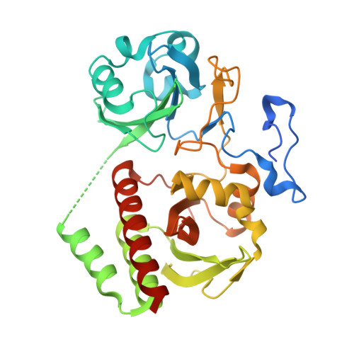
Conserved histidine and tyrosine determine spectral responses through the water network in Deinococcus radiodurans phytochrome.

Lehtivuori, H.Rumfeldt, J.Mustalahti, S.Kurkinen, S.Takala, H.

(2022) Photochem Photobiol Sci 21: 1975-1989

  • DOI: https://doi.org/10.1007/s43630-022-00272-6
  • Primary Citation of Related Structures:  
    7Z9D, 7Z9E

  • PubMed Abstract: 

    Phytochromes are red light-sensing photoreceptor proteins that bind a bilin chromophore. Here, we investigate the role of a conserved histidine (H260) and tyrosine (Y263) in the chromophore-binding domain (CBD) of Deinococcus radiodurans phytochrome (DrBphP). Using crystallography, we show that in the H260A variant, the missing imidazole side chain leads to increased water content in the binding pocket. On the other hand, Y263F mutation reduces the water occupancy around the chromophore. Together, these changes in water coordination alter the protonation and spectroscopic properties of the biliverdin. These results pinpoint the importance of this conserved histidine and tyrosine, and the related water network, for the function and applications of phytochromes.


  • Organizational Affiliation

    Nanoscience Center, Department of Physics, University of Jyvaskyla, 40014, Jyvaskyla, Finland.


Macromolecules
Find similar proteins by:  (by identity cutoff)  |  3D Structure
Entity ID: 1
MoleculeChains Sequence LengthOrganismDetailsImage
Bacteriophytochrome343Deinococcus radiodurans R1 = ATCC 13939 = DSM 20539Mutation(s): 2 
Gene Names: bphPDR_A0050
EC: 2.7.13.3
UniProt
Find proteins for Q9RZA4 (Deinococcus radiodurans (strain ATCC 13939 / DSM 20539 / JCM 16871 / CCUG 27074 / LMG 4051 / NBRC 15346 / NCIMB 9279 / VKM B-1422 / R1))
Explore Q9RZA4 
Go to UniProtKB:  Q9RZA4
Entity Groups  
Sequence Clusters30% Identity50% Identity70% Identity90% Identity95% Identity100% Identity
UniProt GroupQ9RZA4
Sequence Annotations
Expand
  • Reference Sequence
Small Molecules
Ligands 3 Unique
IDChains Name / Formula / InChI Key2D Diagram3D Interactions
LBV (Subject of Investigation/LOI)
Query on LBV

Download Ideal Coordinates CCD File 
G [auth A]3-[2-[(Z)-[3-(2-carboxyethyl)-5-[(Z)-(4-ethenyl-3-methyl-5-oxidanylidene-pyrrol-2-ylidene)methyl]-4-methyl-pyrrol-1-ium -2-ylidene]methyl]-5-[(Z)-[(3E)-3-ethylidene-4-methyl-5-oxidanylidene-pyrrolidin-2-ylidene]methyl]-4-methyl-1H-pyrrol-3- yl]propanoic acid
C33 H37 N4 O6
DKMLMZVDTGOEGU-ISEYCTJISA-O
MPD
Query on MPD

Download Ideal Coordinates CCD File 
C [auth A],
F [auth A]
(4S)-2-METHYL-2,4-PENTANEDIOL
C6 H14 O2
SVTBMSDMJJWYQN-YFKPBYRVSA-N
ACT
Query on ACT

Download Ideal Coordinates CCD File 
B [auth A],
D [auth A],
E [auth A]
ACETATE ION
C2 H3 O2
QTBSBXVTEAMEQO-UHFFFAOYSA-M
Experimental Data & Validation

Experimental Data

  • Method: X-RAY DIFFRACTION
  • Resolution: 1.48 Å
  • R-Value Free: 0.169 
  • R-Value Work: 0.128 
  • R-Value Observed: 0.130 
  • Space Group: C 1 2 1
Unit Cell:
Length ( Å )Angle ( ˚ )
a = 93.76α = 90
b = 54.33β = 92.16
c = 70.37γ = 90
Software Package:
Software NamePurpose
XSCALEdata scaling
PHASERphasing
REFMACrefinement
PDB_EXTRACTdata extraction
XDSdata reduction

Structure Validation

View Full Validation Report


Ligand Structure Quality Assessment 


Entry History & Funding Information

Deposition Data

  • Released Date: 2022-08-10 
  • Deposition Author(s): Takala, H.

Funding OrganizationLocationGrant Number
Academy of FinlandFinland285461, 330678

Revision History  (Full details and data files)

  • Version 1.0: 2022-08-10
    Type: Initial release
  • Version 1.1: 2022-11-16
    Changes: Database references
  • Version 1.2: 2024-01-31
    Changes: Data collection, Refinement description