7YW2

Crystal structure of tRNA 2'-phosphotransferase from Mus musculus


Experimental Data Snapshot

  • Method: X-RAY DIFFRACTION
  • Resolution: 2.23 Å
  • R-Value Free: 0.223 
  • R-Value Work: 0.202 
  • R-Value Observed: 0.203 

wwPDB Validation   3D Report Full Report


Ligand Structure Quality Assessment 


This is version 1.1 of the entry. See complete history


Literature

Structural and biochemical insights into the molecular mechanism of TRPT1 for nucleic acid ADP-ribosylation.

Yang, X.Wang, J.Li, S.Li, X.Gong, J.Yan, Z.Zhou, H.Wu, C.Liu, X.

(2023) Nucleic Acids Res 51: 7649-7665

  • DOI: https://doi.org/10.1093/nar/gkad525
  • Primary Citation of Related Structures:  
    7YW2, 7YW3, 7YW4

  • PubMed Abstract: 

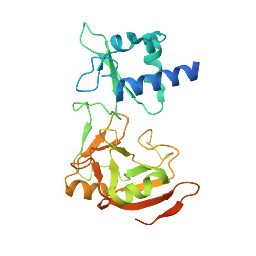
    Nucleic acid ADP-ribosylation has been established as a novel modification found in a wide diversity of prokaryotic and eukaryotic organisms. tRNA 2'-phosphotransferase 1 (TRPT1/TPT1/KptA) possesses ADP-ribosyltransferase (ART) activity and is able to ADP-ribosylate nucleic acids. However, the underlying molecular mechanism remains elusive. Here, we determined crystal structures of TRPT1s in complex with NAD+ from Homo sapiens, Mus musculus and Saccharomyces cerevisiae. Our results revealed that the eukaryotic TRPT1s adopt common mechanisms for both NAD+ and nucleic acid substrate binding. The conserved SGR motif induces a significant conformational change in the donor loop upon NAD+ binding to facilitate the catalytic reaction of ART. Moreover, the nucleic acid-binding residue redundancy provides structural flexibility to accommodate different nucleic acid substrates. Mutational assays revealed that TRPT1s employ different catalytic and nucleic acid-binding residues to perform nucleic acid ADP-ribosylation and RNA 2'-phosphotransferase activities. Finally, cellular assays revealed that the mammalian TRPT1 is able to promote endocervical HeLa cell survival and proliferation. Together, our results provide structural and biochemical insights into the molecular mechanism of TRPT1 for nucleic acid ADP-ribosylation.


  • Organizational Affiliation

    College of Life Sciences, Hebei Innovation Center for Bioengineering and Biotechnology, Institute of Life Sciences and Green Development, Hebei University, Baoding 071002, Hebei, China.


Macromolecules
Find similar proteins by:  (by identity cutoff)  |  3D Structure
Entity ID: 1
MoleculeChains Sequence LengthOrganismDetailsImage
tRNA 2'-phosphotransferase 1249Mus musculusMutation(s): 0 
Gene Names: Trpt1Tpt1h
EC: 2.7.1.160
UniProt
Find proteins for Q8K3A2 (Mus musculus)
Explore Q8K3A2 
Go to UniProtKB:  Q8K3A2
Entity Groups  
Sequence Clusters30% Identity50% Identity70% Identity90% Identity95% Identity100% Identity
UniProt GroupQ8K3A2
Sequence Annotations
Expand
  • Reference Sequence
Small Molecules
Ligands 6 Unique
IDChains Name / Formula / InChI Key2D Diagram3D Interactions
HQG (Subject of Investigation/LOI)
Query on HQG

Download Ideal Coordinates CCD File 
B [auth A][[(2~{R},3~{S},4~{R},5~{R})-5-(6-aminopurin-9-yl)-3,4-bis(oxidanyl)oxolan-2-yl]methoxy-oxidanyl-phosphoryl] [(2~{R},3~{S},4~{R},5~{R})-3,4-bis(oxidanyl)-5-phosphonooxy-oxolan-2-yl]methyl hydrogen phosphate
C15 H24 N5 O17 P3
CUNFRFHBHMFVPH-KEOHHSTQSA-N
EPE (Subject of Investigation/LOI)
Query on EPE

Download Ideal Coordinates CCD File 
E [auth A]4-(2-HYDROXYETHYL)-1-PIPERAZINE ETHANESULFONIC ACID
C8 H18 N2 O4 S
JKMHFZQWWAIEOD-UHFFFAOYSA-N
SOR (Subject of Investigation/LOI)
Query on SOR

Download Ideal Coordinates CCD File 
O [auth A],
P [auth A]
sorbitol
C6 H14 O6
FBPFZTCFMRRESA-JGWLITMVSA-N
PO4 (Subject of Investigation/LOI)
Query on PO4

Download Ideal Coordinates CCD File 
D [auth A]PHOSPHATE ION
O4 P
NBIIXXVUZAFLBC-UHFFFAOYSA-K
GOL (Subject of Investigation/LOI)
Query on GOL

Download Ideal Coordinates CCD File 
C [auth A],
F [auth A],
G [auth A],
N [auth A]
GLYCEROL
C3 H8 O3
PEDCQBHIVMGVHV-UHFFFAOYSA-N
GLY (Subject of Investigation/LOI)
Query on GLY

Download Ideal Coordinates CCD File 
H [auth A]
I [auth A]
J [auth A]
K [auth A]
L [auth A]
H [auth A],
I [auth A],
J [auth A],
K [auth A],
L [auth A],
M [auth A],
Q [auth A],
R [auth A],
S [auth A],
T [auth A]
GLYCINE
C2 H5 N O2
DHMQDGOQFOQNFH-UHFFFAOYSA-N
Experimental Data & Validation

Experimental Data

  • Method: X-RAY DIFFRACTION
  • Resolution: 2.23 Å
  • R-Value Free: 0.223 
  • R-Value Work: 0.202 
  • R-Value Observed: 0.203 
  • Space Group: P 31 2 1
Unit Cell:
Length ( Å )Angle ( ˚ )
a = 65.585α = 90
b = 65.585β = 90
c = 148.459γ = 120
Software Package:
Software NamePurpose
Aimlessdata scaling
PHENIXrefinement
PDB_EXTRACTdata extraction
XDSdata reduction
PHENIXphasing

Structure Validation

View Full Validation Report


Ligand Structure Quality Assessment 


Entry History & Funding Information

Deposition Data


Funding OrganizationLocationGrant Number
National Natural Science Foundation of China (NSFC)China--

Revision History  (Full details and data files)

  • Version 1.0: 2023-07-26
    Type: Initial release
  • Version 1.1: 2023-08-30
    Changes: Data collection, Database references