Mechanism-Guided Computational Design of omega-Transaminase by Reprograming of High-Energy-Barrier Steps.
Yang, L., Zhang, K., Xu, M., Xie, Y., Meng, X., Wang, H., Wei, D.(2022) Angew Chem Int Ed Engl 61: e202212555-e202212555
- PubMed: 36300723 Search on PubMed
- DOI: https://doi.org/10.1002/anie.202212555
- Primary Citation Related Structures:
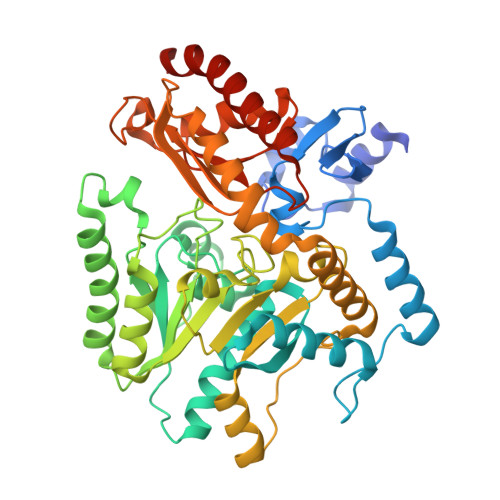
7YPM, 7YPN - PubMed Abstract:
ω-Transaminases (ω-TAs) show considerable potential for the synthesis of chiral amines. However, their low catalytic efficiency towards bulky substrates limits their application, and complicated catalytic mechanisms prevent precise enzyme design. Herein, we address this challenge using a mechanism-guided computational enzyme design strategy by reprograming the transition and ground states in key reaction steps. The common features among the three high-energy-barrier steps responsible for the low catalytic efficiency were revealed using quantum mechanics (QM). Five key residues were simultaneously tailored to stabilize the rate-limiting transition state with the aid of the Rosetta design. The 14 top-ranked variants showed 16.9-143-fold improved catalytic activity. The catalytic efficiency of the best variant, M9 (Q25F/M60W/W64F/I266A), was significantly increased, with a 1660-fold increase in k cat /K m and a 1.5-26.8-fold increase in turnover number (TON) towards various indanone derivatives.
- State Key Laboratory of Bioreactor Engineering, New World Institute of Biotechnology East China University of Science and Technology, Shanghai, P. R. China.
Organizational Affiliation: