7YOB

Crystal structure of Aldehyde dehydrogenase 1A1 from mouse


Experimental Data Snapshot

  • Method: X-RAY DIFFRACTION
  • Resolution: 2.89 Å
  • R-Value Free: 0.249 
  • R-Value Work: 0.195 
  • R-Value Observed: 0.196 

wwPDB Validation   3D Report Full Report


This is version 2.2 of the entry. See complete history


Literature

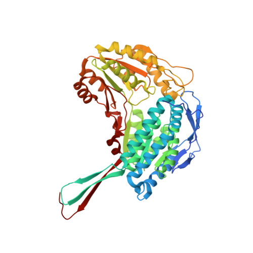
Crystal structure of aldehyde dehydrogenase 1A1 from mouse.

Zhang, X.Ouyang, Z.

(2022) Biochem Biophys Res Commun 628: 141-146

  • DOI: https://doi.org/10.1016/j.bbrc.2022.08.054
  • Primary Citation of Related Structures:  
    7YOB

  • PubMed Abstract: 

    Aldehyde dehydrogenase 1A1 (ALDH1A1) is an enzyme that catalyzes the NAD + -dependent oxidation of aldehydes to carboxylic acids, participating in various metabolic processes. Currently, only structures from human and Ovis aries have been reported. Here we show a 2.89 Å resolution structure of ALDH1A1 from mice using X-ray crystallography. We performed a detailed analysis of the structure and compared it with ALDH1A1 structures from two other species, highlighting the significance of the differences. Structural superimposition reveals that the tetrameric molecule is asymmetrical, and the NAD + -binding domain exhibits a certain rotation. In addition, the noticeable structural differences were detected, including the unique contact between Ser461 and Asp148, as well as the side chain orientations of three amino acids residues, Asn474, Met471 and Phe466. This study helps to expand the structural diversity of the ALDH family.


  • Organizational Affiliation

    Department of Pathogen Biology, School of Basic Medicine, Tongji Medical College, Huazhong University of Science and Technology, 13 Hangkong Road, Wuhan, Hubei, 430030, China.


Macromolecules
Find similar proteins by:  (by identity cutoff)  |  3D Structure
Entity ID: 1
MoleculeChains Sequence LengthOrganismDetailsImage
Aldehyde dehydrogenase 1A1507Mus musculusMutation(s): 0 
Gene Names: Aldh1a1Ahd-2Ahd2Aldh1
EC: 1.2.1.19 (PDB Primary Data), 1.2.1.28 (PDB Primary Data), 1.2.1.3 (PDB Primary Data), 1.2.1.36 (PDB Primary Data)
UniProt
Find proteins for P24549 (Mus musculus)
Explore P24549 
Go to UniProtKB:  P24549
Entity Groups  
Sequence Clusters30% Identity50% Identity70% Identity90% Identity95% Identity100% Identity
UniProt GroupP24549
Sequence Annotations
Expand
  • Reference Sequence
Experimental Data & Validation

Experimental Data

  • Method: X-RAY DIFFRACTION
  • Resolution: 2.89 Å
  • R-Value Free: 0.249 
  • R-Value Work: 0.195 
  • R-Value Observed: 0.196 
  • Space Group: P 1 21 1
Unit Cell:
Length ( Å )Angle ( ˚ )
a = 104.062α = 90
b = 156.484β = 97.97
c = 129.209γ = 90
Software Package:
Software NamePurpose
PHENIXrefinement
XDSdata reduction
Aimlessdata scaling
PHASERphasing

Structure Validation

View Full Validation Report


Entry History & Funding Information

Deposition Data


Funding OrganizationLocationGrant Number
National Natural Science Foundation of China (NSFC)China31870751

Revision History  (Full details and data files)

  • Version 1.0: 2022-08-17
    Type: Initial release
  • Version 2.0: 2022-08-31
    Type: Coordinate replacement
    Reason: Model orientation/position
    Changes: Advisory, Atomic model, Data collection, Database references, Derived calculations, Other, Polymer sequence, Refinement description, Structure summary
  • Version 2.1: 2023-03-01
    Changes: Database references
  • Version 2.2: 2023-11-29
    Changes: Data collection, Refinement description