


Crystal structure of human cytosolic beta-alanyl lysine dipeptidase (PM20D2)
Chandravanshi, K., Gaur, N.K., Kumar, A., Makde, R.D.To be published.
Experimental Data Snapshot
Starting Model: experimental
View more details
wwPDB Validation 3D Report Full Report

Entity ID: 1 | |||||
|---|---|---|---|---|---|
| Molecule | Chains | Sequence Length | Organism | Details | Image |
| Xaa-Arg dipeptidase | 454 | Homo sapiens | Mutation(s): 0 Gene Names: PM20D2, ACY1L2 EC: 3.4.13.4 | 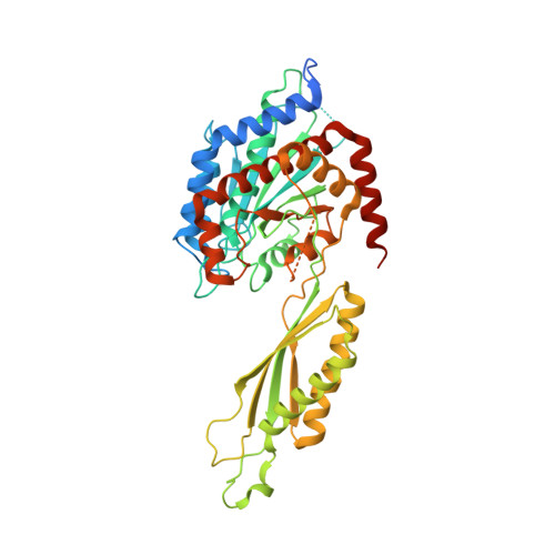 | |
UniProt & NIH Common Fund Data Resources | |||||
PHAROS: Q8IYS1 GTEx: ENSG00000146281 | |||||
Entity Groups | |||||
| Sequence Clusters | 30% Identity50% Identity70% Identity90% Identity95% Identity100% Identity | ||||
| UniProt Group | Q8IYS1 | ||||
Sequence AnnotationsExpand | |||||
Reference Sequence | |||||
| Ligands 1 Unique | |||||
|---|---|---|---|---|---|
| ID | Chains | Name / Formula / InChI Key | 2D Diagram | 3D Interactions | |
| ZN (Subject of Investigation/LOI) Download:Ideal Coordinates CCD File | B [auth A], C [auth A] | ZINC ION Zn PTFCDOFLOPIGGS-UHFFFAOYSA-N |  | ||
| Length ( Å ) | Angle ( ˚ ) |
|---|---|
| a = 78.834 | α = 90 |
| b = 112.888 | β = 90 |
| c = 123.217 | γ = 90 |
| Software Name | Purpose |
|---|---|
| PHENIX | refinement |
| PDB_EXTRACT | data extraction |
| XDS | data reduction |
| Aimless | data scaling |
| PHASER | phasing |
| Funding Organization | Location | Grant Number |
|---|---|---|
| Department of Science & Technology (DST, India) | India | -- |


