7YEU

Superfolder green fluorescent protein with phosphine unnatural amino acid P3BF


Experimental Data Snapshot

  • Method: X-RAY DIFFRACTION
  • Resolution: 1.95 Å
  • R-Value Free: 0.191 
  • R-Value Work: 0.160 

wwPDB Validation   3D Report Full Report


This is version 2.1 of the entry. See complete history


Literature

Genetically Encoded Phosphine Ligand for Metalloprotein Design.

Duan, H.Z.Hu, C.Li, Y.L.Wang, S.H.Xia, Y.Liu, X.Wang, J.Chen, Y.X.

(2022) J Am Chem Soc 144: 22831-22837

  • DOI: https://doi.org/10.1021/jacs.2c09683
  • Primary Citation of Related Structures:  
    7YEU

  • PubMed Abstract: 

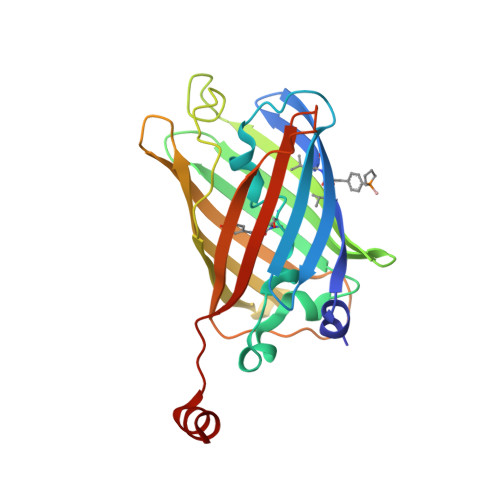
    Phosphine ligands are the most important class of ligands for cross-coupling reactions due to their unique electronic and steric properties. However, metalloproteins generally rely on nitrogen, sulfur, or oxygen ligands. Here, we report the genetic incorporation of P3BF, which contains a biocompatible borane-protected phosphine, into proteins. This step is followed by a straightforward one-pot strategy to perform deboronation and palladium coordination in aqueous and aerobic conditions. The genetically encoded phosphine ligand P3BF should significantly expand our ability to design functional metalloproteins.


  • Organizational Affiliation

    Key Laboratory of Bioorganic Phosphorus Chemistry and Chemical Biology (Ministry of Education), Department of Chemistry, Tsinghua University, Beijing 100084, P.R. China.


Macromolecules
Find similar proteins by:  (by identity cutoff)  |  3D Structure
Entity ID: 1
MoleculeChains Sequence LengthOrganismDetailsImage
Superfolder green fluorescent protein240Aequorea victoriaMutation(s): 0 
UniProt
Find proteins for P42212 (Aequorea victoria)
Explore P42212 
Go to UniProtKB:  P42212
Entity Groups  
Sequence Clusters30% Identity50% Identity70% Identity90% Identity95% Identity100% Identity
UniProt GroupP42212
Sequence Annotations
Expand
  • Reference Sequence
Small Molecules
Modified Residues  1 Unique
IDChains TypeFormula2D DiagramParent
GYS
Query on GYS
A
L-PEPTIDE LINKINGC14 H15 N3 O5SER, TYR, GLY
Experimental Data & Validation

Experimental Data

  • Method: X-RAY DIFFRACTION
  • Resolution: 1.95 Å
  • R-Value Free: 0.191 
  • R-Value Work: 0.160 
  • Space Group: P 41 21 2
Unit Cell:
Length ( Å )Angle ( ˚ )
a = 87.603α = 90
b = 87.603β = 90
c = 114.571γ = 90
Software Package:
Software NamePurpose
REFMACrefinement
PHASERphasing
Aimlessdata scaling
XDSdata reduction

Structure Validation

View Full Validation Report


Entry History & Funding Information

Deposition Data


Funding OrganizationLocationGrant Number
National Natural Science Foundation of China (NSFC)China22177059
Ministry of Science and Technology (MoST, China)China2021YFC2103900
Ministry of Science and Technology (MoST, China)China2018YFA0507600

Revision History  (Full details and data files)

  • Version 1.0: 2023-07-26
    Type: Initial release
  • Version 2.0: 2023-11-15
    Changes: Atomic model, Data collection, Derived calculations
  • Version 2.1: 2024-02-28
    Changes: Database references, Structure summary