7Y6L | pdb_00007y6l

The SARS-CoV-2 receptor binding domain bound with the Fab fragment of a human neutralizing antibody Ab816


Experimental Data Snapshot

  • Method: ELECTRON MICROSCOPY
  • Resolution: 3.50 Å
  • Aggregation State: PARTICLE 
  • Reconstruction Method: SINGLE PARTICLE 

Starting Model: experimental
View more details

wwPDB Validation 3D Report Full Report

Validation slider image for 7Y6L

This is version 1.2 of the entry. See complete history

Literature

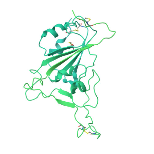
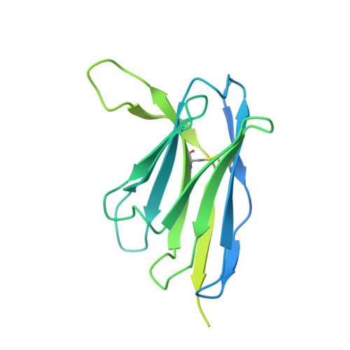
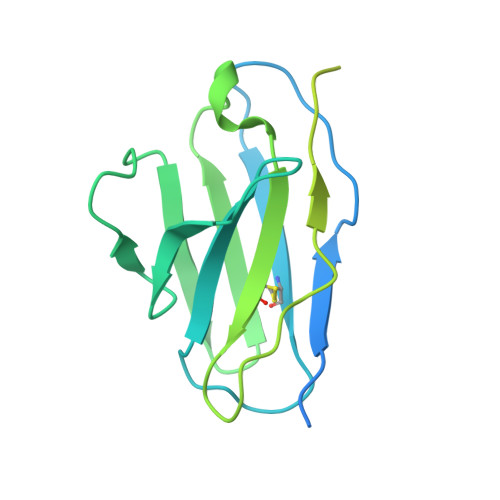
Potent neutralizing broad-spectrum antibody against SARS-CoV-2 generated from dual-antigen-specific B cells from convalescents.

Takeshita, M.Fukuyama, H.Kamada, K.Matsumoto, T.Makino-Okamura, C.Lin, Q.Sakuma, M.Kawahara, E.Yamazaki, I.Uchikubo-Kamo, T.Tomabechi, Y.Hanada, K.Hisano, T.Moriyama, S.Takahashi, Y.Ito, M.Imai, M.Maemura, T.Furusawa, Y.Yamayoshi, S.Kawaoka, Y.Shirouzu, M.Ishii, M.Saya, H.Kondo, Y.Kaneko, Y.Suzuki, K.Fukunaga, K.Takeuchi, T.

(2023) iScience 26: 106955-106955

  • DOI: https://doi.org/10.1016/j.isci.2023.106955
  • Primary Citation Related Structures: 
    7X93, 7X94, 7X95, 7X96, 7Y6L, 7Y6N

  • PubMed Abstract: 

    Several antibody therapeutics have been developed against SARS-CoV-2; however, they have attenuated neutralizing ability against variants. In this study, we generated multiple broadly neutralizing antibodies from B cells of convalescents, by using two types of receptor-binding domains, Wuhan strain and the Gamma variant as bait. From 172 antibodies generated, six antibodies neutralized all strains prior to the Omicron variant, and the five antibodies were able to neutralize some of the Omicron sub-strains. Structural analysis showed that these antibodies have a variety of characteristic binding modes, such as ACE2 mimicry. We subjected a representative antibody to the hamster infection model after introduction of the N297A modification, and observed a dose-dependent reduction of the lung viral titer, even at a dose of 2 mg/kg. These results demonstrated that our antibodies have certain antiviral activity as therapeutics, and highlighted the importance of initial cell-screening strategy for the efficient development of therapeutic antibodies.


  • Organizational Affiliation
    • Division of Rheumatology, Department of Internal Medicine, Keio University School of Medicine, Tokyo 160-8582, Japan.

Macromolecule Content 

  • Total Structure Weight: 196.76 kDa 
  • Atom Count: 3,308 
  • Modeled Residue Count: 422 
  • Deposited Residue Count: 1,780 
  • Unique protein chains: 3

Macromolecules

Find similar proteins by:|  3D Structure
Entity ID: 1
MoleculeChains  Sequence LengthOrganismDetailsImage
Spike glycoprotein1,278Severe acute respiratory syndrome coronavirus 2Mutation(s): 9 
Gene Names: S2
UniProt
Find proteins for P0DTC2 (Severe acute respiratory syndrome coronavirus 2)
Explore P0DTC2 
Go to UniProtKB:  P0DTC2
Entity Groups
Sequence Clusters30% Identity50% Identity70% Identity90% Identity95% Identity100% Identity
UniProt GroupP0DTC2
Glycosylation
Glycosylation Sites: 1Go to GlyGen: P0DTC2-1
Sequence Annotations
Expand
Reference Sequence
Find similar proteins by:|  3D Structure
Entity ID: 2
MoleculeChains  Sequence LengthOrganismDetailsImage
Ab816 heavy chainB [auth H]264Homo sapiensMutation(s): 0 
Find similar proteins by:|  3D Structure
Entity ID: 3
MoleculeChains  Sequence LengthOrganismDetailsImage
Ab816 light chainC [auth L]238Homo sapiensMutation(s): 0 

Small Molecules

Ligands 1 Unique
IDChains Name / Formula / InChI Key2D Diagram3D Interactions
NAG

Query on NAG


Download:Ideal Coordinates CCD File
D [auth A]2-acetamido-2-deoxy-beta-D-glucopyranose
C8 H15 N O6
OVRNDRQMDRJTHS-FMDGEEDCSA-N

Experimental Data & Validation

Experimental Data

  • Method: ELECTRON MICROSCOPY
  • Resolution: 3.50 Å
  • Aggregation State: PARTICLE 
  • Reconstruction Method: SINGLE PARTICLE 
EM Software:
TaskSoftware PackageVersion
MODEL REFINEMENTPHENIX1.19.2-4158
RECONSTRUCTIONRELION3.1.3

Structure Validation

View Full Validation Report


Entry History 

& Funding Information

Deposition Data


Funding OrganizationLocationGrant Number
Japan Agency for Medical Research and Development (AMED)JapanJP21fk0108468

Revision History  (Full details and data files)

  • Version 1.0: 2023-06-21
    Type: Initial release
  • Version 1.1: 2023-09-13
    Changes: Data collection, Database references
  • Version 1.2: 2024-11-13
    Changes: Data collection, Refinement description, Structure summary