7Y6K | pdb_00007y6k

Cryo-EM structure of SARS-CoV-2 receptor binding domain in complex with K202.B bispecific antibody


Experimental Data Snapshot

  • Method: ELECTRON MICROSCOPY
  • Resolution: 3.34 Å
  • Aggregation State: PARTICLE 
  • Reconstruction Method: SINGLE PARTICLE 

Starting Models: experimental
View more details

wwPDB Validation 3D Report Full Report

Validation slider image for 7Y6K

This is version 3.1 of the entry. See complete history

Literature

Novel bispecific human antibody platform specifically targeting a fully open spike conformation potently neutralizes multiple SARS-CoV-2 variants.

Kim, J.W.Heo, K.Kim, H.J.Yoo, Y.Cho, H.S.Jang, H.J.Lee, H.Y.Ko, I.Y.Woo, J.R.Cho, Y.B.Lee, J.H.Yang, H.R.Shin, H.G.Choi, H.L.Hwang, K.Kim, S.Kim, H.Chun, K.Lee, S.

(2023) Antiviral Res 212: 105576-105576

  • DOI: https://doi.org/10.1016/j.antiviral.2023.105576
  • Primary Citation Related Structures: 
    7Y6K

  • PubMed Abstract: 

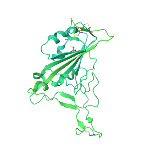
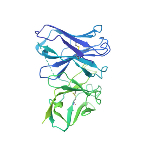
    Rapid emergence of new variants of severe acute respiratory syndrome coronavirus 2 (SARS-CoV-2) has prompted an urgent need for the development of broadly applicable and potently neutralizing antibody platform against the SARS-CoV-2, which can be used for combatting the coronavirus disease 2019 (COVID-19). In this study, based on a noncompeting pair of phage display-derived human monoclonal antibodies (mAbs) specific to the receptor-binding domain (RBD) of SARS-CoV-2 isolated from human synthetic antibody library, we generated K202.B, a novel engineered bispecific antibody with an immunoglobulin G4-single-chain variable fragment design, with sub- or low nanomolar antigen-binding avidity. Compared with the parental mAbs or mAb cocktail, the K202.B antibody showed superior neutralizing potential against a variety of SARS-CoV-2 variants in vitro. Furthermore, structural analysis of bispecific antibody-antigen complexes using cryo-electron microscopy revealed the mode of action of K202.B complexed with a fully open three-RBD-up conformation of SARS-CoV-2 trimeric spike proteins by simultaneously interconnecting two independent epitopes of the SARS-CoV-2 RBD via inter-protomer interactions. Intravenous monotherapy using K202.B exhibited potent neutralizing activity in SARS-CoV-2 wild-type- and B.1.617.2 variant-infected mouse models, without significant toxicity in vivo. The results indicate that this novel approach of development of immunoglobulin G4-based bispecific antibody from an established human recombinant antibody library is likely to be an effective strategy for the rapid development of bispecific antibodies, and timely management against fast-evolving SARS-CoV-2 variants.


  • Organizational Affiliation
    • Department of Biochemistry, Kookmin University, Seoul, 02707, Republic of Korea.

Macromolecule Content 

  • Total Structure Weight: 233.5 kDa 
  • Atom Count: 6,363 
  • Modeled Residue Count: 834 
  • Deposited Residue Count: 2,140 
  • Unique protein chains: 3

Macromolecules

Find similar proteins by:|  3D Structure
Entity ID: 1
MoleculeChains  Sequence LengthOrganismDetailsImage
Spike glycoproteinA [auth B]1,208Severe acute respiratory syndrome coronavirus 2Mutation(s): 9 
Gene Names: S2
UniProt
Find proteins for P0DTC2 (Severe acute respiratory syndrome coronavirus 2)
Explore P0DTC2 
Go to UniProtKB:  P0DTC2
Entity Groups
Sequence Clusters30% Identity50% Identity70% Identity90% Identity95% Identity100% Identity
UniProt GroupP0DTC2
Glycosylation
Glycosylation Sites: 1Go to GlyGen: P0DTC2-1
Sequence Annotations
Expand
Reference Sequence
Find similar proteins by:|  3D Structure
Entity ID: 2
MoleculeChains  Sequence LengthOrganismDetailsImage
Bispecific anitybody (scFv and light chain of the antibody)B [auth E]478Homo sapiensMutation(s): 0 
Find similar proteins by:|  3D Structure
Entity ID: 3
MoleculeChains  Sequence LengthOrganismDetailsImage
Heavy chain from K202.B antibodyC [auth J]454Homo sapiensMutation(s): 0 

Small Molecules

Ligands 1 Unique
IDChains Name / Formula / InChI Key2D Diagram3D Interactions
NAG

Query on NAG


Download:Ideal Coordinates CCD File
D [auth B]2-acetamido-2-deoxy-beta-D-glucopyranose
C8 H15 N O6
OVRNDRQMDRJTHS-FMDGEEDCSA-N

Experimental Data & Validation

Experimental Data

  • Method: ELECTRON MICROSCOPY
  • Resolution: 3.34 Å
  • Aggregation State: PARTICLE 
  • Reconstruction Method: SINGLE PARTICLE 
EM Software:
TaskSoftware PackageVersion
RECONSTRUCTIONcryoSPARC
MODEL REFINEMENTPHENIX1.20.1
MODEL REFINEMENTCoot0.9.8.1

Structure Validation

View Full Validation Report


Entry History 

& Funding Information

Deposition Data


Funding OrganizationLocationGrant Number
National Research Foundation (NRF, Korea)Korea, Republic Of--

Revision History  (Full details and data files)

  • Version 1.0: 2023-07-19
    Type: Initial release
  • Version 1.1: 2024-10-30
    Changes: Data collection, Refinement description, Structure summary
  • Version 2.0: 2025-04-02
    Changes: Advisory, Atomic model, Data collection, Database references, Derived calculations, Polymer sequence, Source and taxonomy, Structure summary
  • Version 3.0: 2025-06-18
    Changes: Advisory, Atomic model, Data collection, Database references, Derived calculations, Polymer sequence, Source and taxonomy, Structure summary
  • Version 3.1: 2026-04-22
    Changes: Data collection, Structure summary