7Y6A | pdb_00007y6a

Crystal structure of Chicken Egg Lysozyme

  • Classification: HYDROLASE
  • Organism(s): Gallus gallus
  • Mutation(s): No 

  • Deposited: 2022-06-18 Released: 2023-04-19 
  • Deposition Author(s): DeMirci, H.
  • Funding Organization(s): Other government

Experimental Data Snapshot

  • Method: X-RAY DIFFRACTION
  • Resolution: 1.50 Å
  • R-Value Free: 
    0.256 (Depositor), 0.254 (DCC) 
  • R-Value Work: 
    0.222 (Depositor), 0.222 (DCC) 
  • R-Value Observed: 
    0.223 (Depositor) 

Starting Model: experimental
View more details

wwPDB Validation 3D Report Full Report

Validation slider image for 7Y6A

This is version 1.3 of the entry. See complete history

Literature

Cryogenic X-ray crystallographic studies of biomacromolecules at Turkish Light Source " Turkish DeLight ".

Atalay, N.Akcan, E.K.Gul, M.Ayan, E.Destan, E.Ertem, F.B.Tokay, N.Cakilkaya, B.Nergiz, Z.Karakadioglu, G.Kepceoglu, A.Yapici, I.Tosun, B.Baldir, N.Yildirim, G.Johnson, J.A.Guven, O.Shafiei, A.Arslan, N.E.Yilmaz, M.Kulakman, C.Paydos, S.S.Cinal, Z.S.Sabanoglu, K.Pazarceviren, A.Yilmaz, A.Canbay, B.Asci, B.Kartal, E.Tavli, S.Caliseki, M.Goc, G.Mermer, A.Yesilay, G.Altuntas, S.Tateishi, H.Otsuka, M.Fujita, M.Tekin, S.Ciftci, H.Durdagi, S.Dinler Doganay, G.Karaca, E.Kaplan Turkoz, B.Kabasakal, B.V.Kati, A.Demirci, H.

(2023) Turk J Biol 47: 1-13

  • DOI: https://doi.org/10.55730/1300-0152.2637
  • Primary Citation Related Structures: 
    7Y6A

  • PubMed Abstract: 

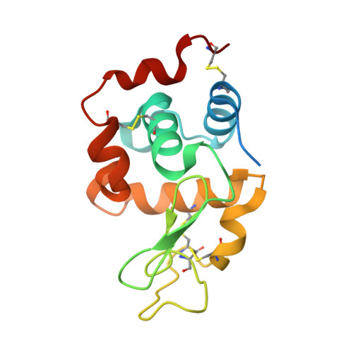
    X-ray crystallography is a robust and powerful structural biology technique that provides high-resolution atomic structures of biomacromolecules. Scientists use this technique to unravel mechanistic and structural details of biological macromolecules (e.g., proteins, nucleic acids, protein complexes, protein-nucleic acid complexes, or large biological compartments). Since its inception, single-crystal cryocrystallography has never been performed in Türkiye due to the lack of a single-crystal X-ray diffractometer. The X-ray diffraction facility recently established at the University of Health Sciences, İstanbul, Türkiye will enable Turkish and international researchers to easily perform high-resolution structural analysis of biomacromolecules from single crystals. Here, we describe the technical and practical outlook of a state-of-the-art home-source X-ray, using lysozyme as a model protein. The methods and practice described in this article can be applied to any biological sample for structural studies. Therefore, this article will be a valuable practical guide from sample preparation to data analysis.


  • Organizational Affiliation
    • Department of Molecular Biology and Genetics, Faculty of Science, Koç University, İstanbul, Türkiye.

Macromolecule Content 

  • Total Structure Weight: 16.32 kDa 
  • Atom Count: 1,140 
  • Modeled Residue Count: 128 
  • Deposited Residue Count: 147 
  • Unique protein chains: 1

Macromolecules

Find similar proteins by:|  3D Structure
Entity ID: 1
MoleculeChains  Sequence LengthOrganismDetailsImage
Lysozyme C147Gallus gallusMutation(s): 0 
EC: 3.2.1.17
UniProt
Find proteins for P00698 (Gallus gallus)
Explore P00698 
Go to UniProtKB:  P00698
Entity Groups
Sequence Clusters30% Identity50% Identity70% Identity90% Identity95% Identity100% Identity
UniProt GroupP00698
Sequence Annotations
Expand
Reference Sequence

Small Molecules

Ligands 1 Unique
IDChains Name / Formula / InChI Key2D Diagram3D Interactions
NO3
(Subject of Investigation/LOI)

Query on NO3


Download:Ideal Coordinates CCD File
B [auth A]NITRATE ION
N O3
NHNBFGGVMKEFGY-UHFFFAOYSA-N

Experimental Data & Validation

Experimental Data

  • Method: X-RAY DIFFRACTION
  • Resolution: 1.50 Å
  • R-Value Free:  0.256 (Depositor), 0.254 (DCC) 
  • R-Value Work:  0.222 (Depositor), 0.222 (DCC) 
  • R-Value Observed: 0.223 (Depositor) 
Space Group: P 43 21 2
Unit Cell:
Length ( Å )Angle ( ˚ )
a = 77.35α = 90
b = 77.35β = 90
c = 38.8γ = 90
Software Package:
Software NamePurpose
CrysalisProdata scaling
PHENIXrefinement
PDB_EXTRACTdata extraction
CrysalisProdata reduction
PHENIXphasing

Structure Validation

View Full Validation Report


Entry History 

& Funding Information

Deposition Data

  • Released Date: 2023-04-19 
  • Deposition Author(s): DeMirci, H.

Funding OrganizationLocationGrant Number
Other governmentTurkey118C270

Revision History  (Full details and data files)

  • Version 1.0: 2023-04-19
    Type: Initial release
  • Version 1.1: 2023-08-16
    Changes: Data collection, Database references
  • Version 1.2: 2023-11-29
    Changes: Refinement description
  • Version 1.3: 2024-11-13
    Changes: Database references, Structure summary