


Crystal structure of DUSP10 mutant_N130A
Hu, I.C., Lyu, P.C.To be published.
Experimental Data Snapshot
Starting Model: experimental
View more details
wwPDB Validation 3D Report Full Report

Entity ID: 1 | |||||
|---|---|---|---|---|---|
| Molecule | Chains | Sequence Length | Organism | Details | Image |
| Dual specificity protein phosphatase 10 | 149 | Homo sapiens | Mutation(s): 1 Gene Names: DUSP10, MKP5 EC: 3.1.3.16 (PDB Primary Data), 3.1.3.48 (PDB Primary Data) | 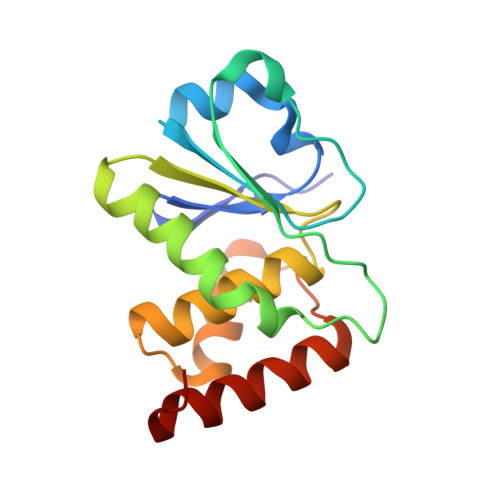 | |
UniProt & NIH Common Fund Data Resources | |||||
PHAROS: Q9Y6W6 GTEx: ENSG00000143507 | |||||
Entity Groups | |||||
| Sequence Clusters | 30% Identity50% Identity70% Identity90% Identity95% Identity100% Identity | ||||
| UniProt Group | Q9Y6W6 | ||||
Sequence AnnotationsExpand | |||||
Reference Sequence | |||||
| Length ( Å ) | Angle ( ˚ ) |
|---|---|
| a = 39.42 | α = 95.76 |
| b = 41.156 | β = 99.68 |
| c = 55.742 | γ = 117.23 |
| Software Name | Purpose |
|---|---|
| PHENIX | refinement |
| HKL-2000 | data reduction |
| HKL-2000 | data scaling |
| HKL-2000 | data collection |
| MOLREP | phasing |
| Funding Organization | Location | Grant Number |
|---|---|---|
| Ministry of Science and Technology (MoST, Taiwan) | Taiwan | -- |


