7Y3J | pdb_00007y3j

24B3 antibody-peptide complex


Experimental Data Snapshot

  • Method: X-RAY DIFFRACTION
  • Resolution: 2.60 Å
  • R-Value Free: 
    0.261 (Depositor), 0.265 (DCC) 
  • R-Value Work: 
    0.222 (Depositor), 0.221 (DCC) 
  • R-Value Observed: 
    0.224 (Depositor) 

Starting Model: experimental
View more details

wwPDB Validation 3D Report Full Report

Validation slider image for 7Y3J

This is version 1.2 of the entry. See complete history

Literature

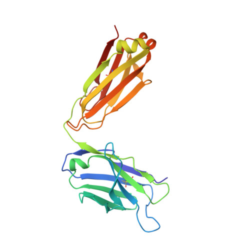
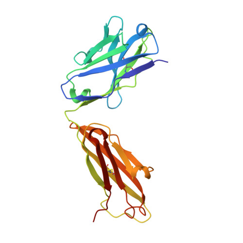
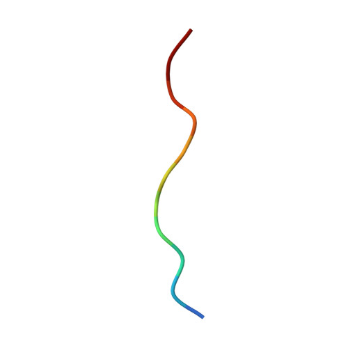
Structural basis of the 24B3 antibody against the toxic conformer of amyloid beta with a turn at positions 22 and 23.

Irie, Y.Matsushima, Y.Kita, A.Miki, K.Segawa, T.Maeda, M.Yanagita, R.C.Irie, K.

(2022) Biochem Biophys Res Commun 621: 162-167

  • DOI: https://doi.org/10.1016/j.bbrc.2022.07.010
  • Primary Citation Related Structures: 
    7Y3J

  • PubMed Abstract: 

    Amyloid β-protein (Aβ) oligomers are involved in the early stages of Alzheimer's disease (AD) and antibodies against these toxic oligomers could be useful for accurate diagnosis of AD. We identified the toxic conformer of Aβ42 with a turn at positions 22/23, which has a propensity to form toxic oligomers. The antibody 24B3, developed by immunization of a toxic conformer surrogate E22P-Aβ9-35 in mice, was found to be useful for AD diagnosis using human cerebrospinal fluid (CSF). However, it is not known how 24B3 recognizes the toxic conformation of wild-type Aβ in CSF. Here, we report the crystal structure of 24B3 Fab complexed with E22P-Aβ11-34, whose residues 16-26 were observed in electron densities, suggesting that the residues comprising the toxic turn at positions 22/23 were recognized by 24B3. Since 24B3 bound only to Aβ42 aggregates, several conformationally restricted analogs of Aβ42 with an intramolecular disulfide bond to mimic the conformation of toxic Aβ42 aggregates were screened by enzyme immunoassay. As a result, only F19C,A30homoC-SS-Aβ42 (1) bound significantly to 24B3. These data provide a structural basis for its low affinity to the Aβ42 monomer and selectivity for its aggregate form.


  • Organizational Affiliation
    • Division of Food Science and Biotechnology, Graduate School of Agriculture, Kyoto University, Sakyo-Ku, Kyoto, 606-8502, Japan.

Macromolecule Content 

  • Total Structure Weight: 48.79 kDa 
  • Atom Count: 3,452 
  • Modeled Residue Count: 447 
  • Deposited Residue Count: 447 
  • Unique protein chains: 3

Macromolecules

Find similar proteins by:|  3D Structure
Entity ID: 1
MoleculeChains  Sequence LengthOrganismDetailsImage
24B3 Light chainA [auth L]217Mus musculusMutation(s): 0 
Find similar proteins by:|  3D Structure
Entity ID: 2
MoleculeChains  Sequence LengthOrganismDetailsImage
24B3 Heavy chainB [auth H]219Mus musculusMutation(s): 0 
Find similar proteins by:  Sequence   |   3D Structure  
Entity ID: 3
MoleculeChains  Sequence LengthOrganismDetailsImage
ALA-LEU-VAL-PHE-PHE-ALA-PRO-ALA-VAL-GLY-SERC [auth A]11Homo sapiensMutation(s): 0 
UniProt & NIH Common Fund Data Resources
Find proteins for P05067 (Homo sapiens)
Explore P05067 
Go to UniProtKB:  P05067
PHAROS:  P05067
GTEx:  ENSG00000142192 
Entity Groups
Sequence Clusters30% Identity50% Identity70% Identity90% Identity95% Identity100% Identity
UniProt GroupP05067
Sequence Annotations
Expand
Reference Sequence

Experimental Data & Validation

Experimental Data

  • Method: X-RAY DIFFRACTION
  • Resolution: 2.60 Å
  • R-Value Free:  0.261 (Depositor), 0.265 (DCC) 
  • R-Value Work:  0.222 (Depositor), 0.221 (DCC) 
  • R-Value Observed: 0.224 (Depositor) 
Space Group: C 2 2 21
Unit Cell:
Length ( Å )Angle ( ˚ )
a = 49.589α = 90
b = 127.883β = 90
c = 160.342γ = 90
Software Package:
Software NamePurpose
PHENIXrefinement
HKL-2000data reduction
SCALEPACKdata scaling
MOLREPphasing

Structure Validation

View Full Validation Report


Entry History 

& Funding Information

Deposition Data


Funding OrganizationLocationGrant Number
Japan Society for the Promotion of Science (JSPS)JapanJP19H00921

Revision History  (Full details and data files)

  • Version 1.0: 2022-08-17
    Type: Initial release
  • Version 1.1: 2023-11-29
    Changes: Data collection, Refinement description
  • Version 1.2: 2024-10-23
    Changes: Structure summary