7XZ4

Crystal structure of ferritin from Setaria italica


Experimental Data Snapshot

  • Method: X-RAY DIFFRACTION
  • Resolution: 2.20 Å
  • R-Value Free: 0.201 
  • R-Value Work: 0.158 
  • R-Value Observed: 0.162 

wwPDB Validation   3D Report Full Report


This is version 1.1 of the entry. See complete history


Literature

Extension Peptide of Plant Ferritin from Setaria italica Presents a Novel Fold.

Wang, W.Wang, Y.Xi, H.Song, Z.Zhang, W.Xie, L.Ma, D.Qin, N.Wang, H.

(2023) J Agric Food Chem 71: 934-943

  • DOI: https://doi.org/10.1021/acs.jafc.2c07595
  • Primary Citation of Related Structures:  
    7XZ4

  • PubMed Abstract: 

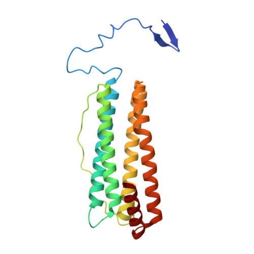
    The extension peptide (EP) is the most distinctive feature of mature plant ferritin. Some EPs have exhibited serine-like protease activity, which is associated with iron uptake and release. EP forms a helix and a long loop, followed by a conserved core helical bundle. However, whether the EP adopts a stable or uniform folding pattern in all plants remains unclear. To clarify this, we investigated the crystal structure of ferritin-1 from Setaria italica (SiFer1), a type of monocotyledon. In our structure of SiFer1, the EP is different from other EPs in other solved structures of plant ferritins and consisted of a pair of β-sheets, a shorter helix, and two loops, which masks two hydrophobic pockets on the outer surface of every subunit. Furthermore, sequence analysis and structure comparison suggest that the EPs in ferritins from monocotyledons may adopt a novel fold pattern, and the conformations of EPs in ferritins are alterable among different plant species. Furthermore, additional eight iron atoms were first founded binding in the fourfold channels, demonstrating the vital function of fourfold channels in iron diffusion. In all, our structure provides new clues for understanding plant ferritins and the functions of the EP.


  • Organizational Affiliation

    Institute of Molecular Science, Shanxi University, Taiyuan 030006, China.


Macromolecules
Find similar proteins by:  (by identity cutoff)  |  3D Structure
Entity ID: 1
MoleculeChains Sequence LengthOrganismDetailsImage
Ferritin208Setaria italicaMutation(s): 0 
Gene Names: 101778084SETIT_8G019400v2
EC: 1.16.3.1
UniProt
Find proteins for K3ZJM0 (Setaria italica)
Explore K3ZJM0 
Go to UniProtKB:  K3ZJM0
Entity Groups  
Sequence Clusters30% Identity50% Identity70% Identity90% Identity95% Identity100% Identity
UniProt GroupK3ZJM0
Sequence Annotations
Expand
  • Reference Sequence
Small Molecules
Ligands 2 Unique
IDChains Name / Formula / InChI Key2D Diagram3D Interactions
PO4
Query on PO4

Download Ideal Coordinates CCD File 
P [auth A],
Q [auth A]
PHOSPHATE ION
O4 P
NBIIXXVUZAFLBC-UHFFFAOYSA-K
FE (Subject of Investigation/LOI)
Query on FE

Download Ideal Coordinates CCD File 
B [auth A]
C [auth A]
D [auth A]
E [auth A]
F [auth A]
B [auth A],
C [auth A],
D [auth A],
E [auth A],
F [auth A],
G [auth A],
H [auth A],
I [auth A],
J [auth A],
K [auth A],
L [auth A],
M [auth A],
N [auth A],
O [auth A]
FE (III) ION
Fe
VTLYFUHAOXGGBS-UHFFFAOYSA-N
Experimental Data & Validation

Experimental Data

  • Method: X-RAY DIFFRACTION
  • Resolution: 2.20 Å
  • R-Value Free: 0.201 
  • R-Value Work: 0.158 
  • R-Value Observed: 0.162 
  • Space Group: I 4 3 2
Unit Cell:
Length ( Å )Angle ( ˚ )
a = 150.336α = 90
b = 150.336β = 90
c = 150.336γ = 90
Software Package:
Software NamePurpose
HKL-2000data reduction
HKL-2000data scaling
PHENIXrefinement
PDB_EXTRACTdata extraction
PHASERphasing

Structure Validation

View Full Validation Report


Entry History & Funding Information

Deposition Data


Funding OrganizationLocationGrant Number
National Natural Science Foundation of China (NSFC)China62075118
National Natural Science Foundation of China (NSFC)China21601112

Revision History  (Full details and data files)

  • Version 1.0: 2023-01-25
    Type: Initial release
  • Version 1.1: 2023-11-29
    Changes: Data collection, Refinement description