7XRL

Diol dehydratase complexed with AdoMeCbl and 1,2-propanediol


Experimental Data Snapshot

  • Method: X-RAY DIFFRACTION
  • Resolution: 1.75 Å
  • R-Value Free: 0.206 
  • R-Value Work: 0.167 
  • R-Value Observed: 0.168 

wwPDB Validation   3D Report Full Report


Ligand Structure Quality Assessment 


This is version 1.1 of the entry. See complete history


Literature

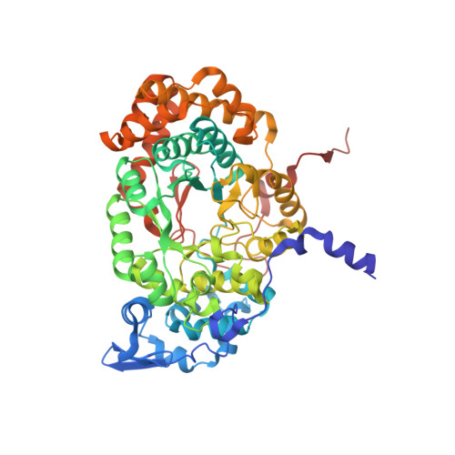
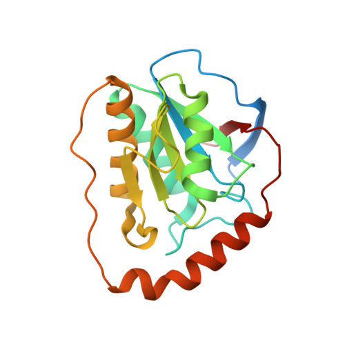
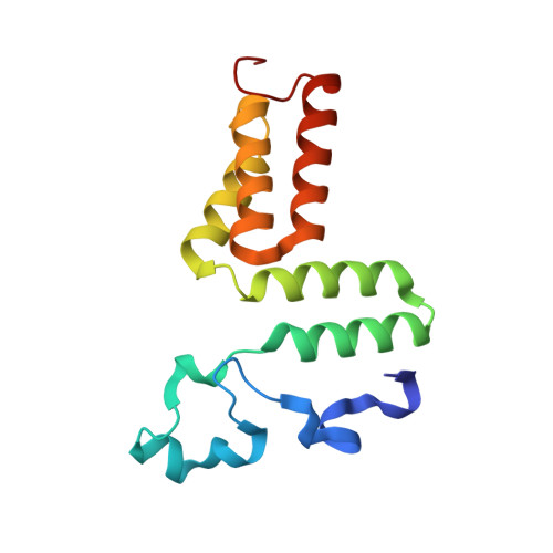
Structural Insights into the Very Low Activity of the Homocoenzyme B 12 Adenosylmethylcobalamin in Coenzyme B 12 -Dependent Diol Dehydratase and Ethanolamine Ammonia-Lyase.

Shibata, N.Higuchi, Y.Krautler, B.Toraya, T.

(2022) Chemistry 28: e202202196-e202202196

  • DOI: https://doi.org/10.1002/chem.202202196
  • Primary Citation of Related Structures:  
    7XRK, 7XRL, 7XRM, 7XRN

  • PubMed Abstract: 

    The X-ray structures of coenzyme B 12 (AdoCbl)-dependent eliminating isomerases complexed with adenosylmethylcobalamin (AdoMeCbl) have been determined. As judged from geometries, the Co-C bond in diol dehydratase (DD) is not activated even in the presence of substrate. In ethanolamine ammonia-lyase (EAL), the bond is elongated in the absence of substrate; in the presence of substrate, the complex likely exists in both pre- and post-homolysis states. The impacts of incorporating an extra CH 2 group are different in the two enzymes: the DD active site is flexible, and AdoMeCbl binding causes large conformational changes that make DD unable to adopt the catalytic state, whereas the EAL active site is rigid, and AdoMeCbl binding does not induce significant conformational changes. Such flexibility and rigidity of the active sites might reflect the tightness of adenine binding. The structures provide good insights into the basis of the very low activity of AdoMeCbl in these enzymes.


  • Organizational Affiliation

    Department of Life Science, Graduate School of Science, University of Hyogo, 3-2-1 Koto, Kamigori-cho, Ako-gun, Hyogo, 678-1297, Japan.


Macromolecules
Find similar proteins by:  (by identity cutoff)  |  3D Structure
Entity ID: 1
MoleculeChains Sequence LengthOrganismDetailsImage
Diol dehydrase alpha subunit
A, D
554Klebsiella oxytocaMutation(s): 0 
Gene Names: pddApduCAB185_12495SAMEA2273575_05741
EC: 4.2.1.28
UniProt
Find proteins for Q59470 (Klebsiella oxytoca)
Explore Q59470 
Go to UniProtKB:  Q59470
Entity Groups  
Sequence Clusters30% Identity50% Identity70% Identity90% Identity95% Identity100% Identity
UniProt GroupQ59470
Sequence Annotations
Expand
  • Reference Sequence
Find similar proteins by:  (by identity cutoff)  |  3D Structure
Entity ID: 2
MoleculeChains Sequence LengthOrganismDetailsImage
Diol dehydrase beta subunit
B, E
200Klebsiella oxytocaMutation(s): 0 
Gene Names: pddB
EC: 4.2.1.28
UniProt
Find proteins for Q59471 (Klebsiella oxytoca)
Explore Q59471 
Go to UniProtKB:  Q59471
Entity Groups  
Sequence Clusters30% Identity50% Identity70% Identity90% Identity95% Identity100% Identity
UniProt GroupQ59471
Sequence Annotations
Expand
  • Reference Sequence
Find similar proteins by:  (by identity cutoff)  |  3D Structure
Entity ID: 3
MoleculeChains Sequence LengthOrganismDetailsImage
Diol dehydrase gamma subunit
C, F
137Klebsiella oxytocaMutation(s): 0 
Gene Names: pddCpduEpduE_1AB185_12485SAMEA2273639_01293SAMEA2273697_01477
EC: 4.2.1.28
UniProt
Find proteins for Q59472 (Klebsiella oxytoca)
Explore Q59472 
Go to UniProtKB:  Q59472
Entity Groups  
Sequence Clusters30% Identity50% Identity70% Identity90% Identity95% Identity100% Identity
UniProt GroupQ59472
Sequence Annotations
Expand
  • Reference Sequence
Small Molecules
Ligands 6 Unique
IDChains Name / Formula / InChI Key2D Diagram3D Interactions
B12 (Subject of Investigation/LOI)
Query on B12

Download Ideal Coordinates CCD File 
L [auth B],
R [auth E]
COBALAMIN
C62 H89 Co N13 O14 P
LKVIQTCSMMVGFU-DWSMJLPVSA-N
FWK (Subject of Investigation/LOI)
Query on FWK

Download Ideal Coordinates CCD File 
K [auth A],
Q [auth D]
(2~{R},3~{R},4~{S},5~{R})-2-(6-aminopurin-9-yl)-5-ethyl-oxolane-3,4-diol
C11 H15 N5 O3
RLOSCLOIJYLPHI-IOSLPCCCSA-N
PGO
Query on PGO

Download Ideal Coordinates CCD File 
G [auth A],
M [auth D]
S-1,2-PROPANEDIOL
C3 H8 O2
DNIAPMSPPWPWGF-VKHMYHEASA-N
CA
Query on CA

Download Ideal Coordinates CCD File 
H [auth A],
N [auth D]
CALCIUM ION
Ca
BHPQYMZQTOCNFJ-UHFFFAOYSA-N
K
Query on K

Download Ideal Coordinates CCD File 
I [auth A],
O [auth D]
POTASSIUM ION
K
NPYPAHLBTDXSSS-UHFFFAOYSA-N
NH4
Query on NH4

Download Ideal Coordinates CCD File 
J [auth A],
P [auth D]
AMMONIUM ION
H4 N
QGZKDVFQNNGYKY-UHFFFAOYSA-O
Experimental Data & Validation

Experimental Data

  • Method: X-RAY DIFFRACTION
  • Resolution: 1.75 Å
  • R-Value Free: 0.206 
  • R-Value Work: 0.167 
  • R-Value Observed: 0.168 
  • Space Group: P 21 21 21
Unit Cell:
Length ( Å )Angle ( ˚ )
a = 73.718α = 90
b = 117.246β = 90
c = 201.55γ = 90
Software Package:
Software NamePurpose
PHENIXrefinement
HKL-2000data reduction
HKL-2000data scaling
PHASERphasing

Structure Validation

View Full Validation Report


Ligand Structure Quality Assessment 


Entry History & Funding Information

Deposition Data


Funding OrganizationLocationGrant Number
Not funded--

Revision History  (Full details and data files)

  • Version 1.0: 2023-03-29
    Type: Initial release
  • Version 1.1: 2023-11-29
    Changes: Data collection, Refinement description