7XQ5

Crystal structure of ScIno2p-ScIno4p bound promoter DNA


Experimental Data Snapshot

  • Method: X-RAY DIFFRACTION
  • Resolution: 2.25 Å
  • R-Value Free: 0.235 
  • R-Value Work: 0.214 
  • R-Value Observed: 0.215 

wwPDB Validation   3D Report Full Report


This is version 1.2 of the entry. See complete history


Literature

Structural Analysis of Ino2p/Ino4p Mutual Interactions and Their Binding Interface with Promoter DNA.

Khan, M.H.Xue, L.Yue, J.Schuller, H.J.Zhu, Z.Niu, L.

(2022) Int J Mol Sci 23

  • DOI: https://doi.org/10.3390/ijms23147600
  • Primary Citation of Related Structures:  
    7XQ5

  • PubMed Abstract: 

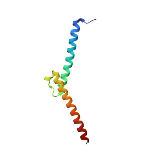
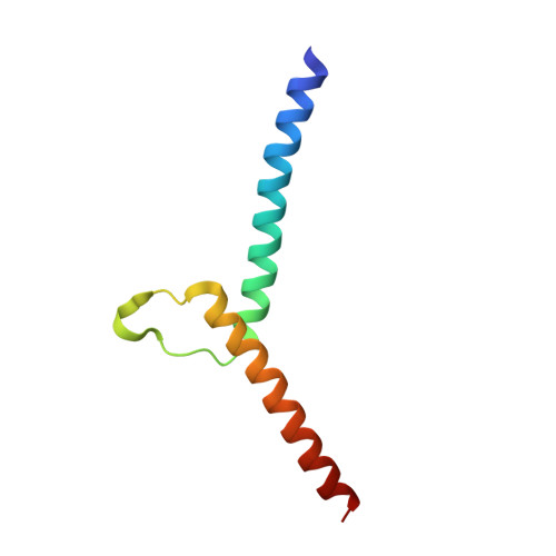
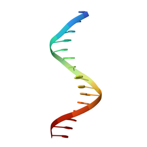
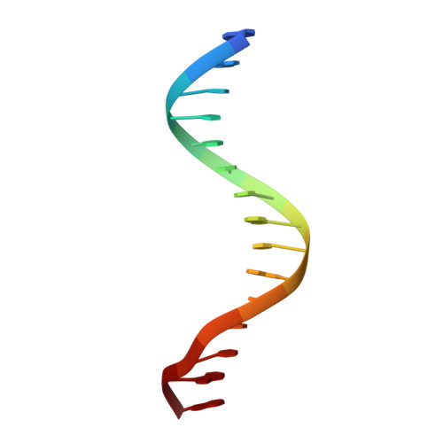
    Gene expression is mediated by a series of regulatory proteins, i.e., transcription factors. Under different growth conditions, the transcriptional regulation of structural genes is associated with the recognition of specific regulatory elements (REs) in promoter DNA. The manner by which transcription factors recognize distinctive REs is a key question in structural biology. Previous research has demonstrated that Ino2p/Ino4p heterodimer is associated with the transcriptional regulation of phospholipid biosynthetic genes. Mechanistically, Ino2p/Ino4p could specifically recognize the inositol/choline-responsive element (ICRE), followed by the transcription activation of the phospholipid biosynthetic gene. While the promoter DNA sequence for Ino2p has already been characterized, the structural basis for the mutual interaction between Ino2p/Ino4p and their binding interface with promoter DNA remain relatively unexplored. Here, we have determined the crystalline structure of the Ino2p DBD /Ino4p DBD /DNA ternary complex, which highlights some residues (Ino2p His12/Glu16/Arg20/Arg44 and Ino4p His12/Glu16/Arg19/Arg20 ) associated with the sequence-specific recognition of promoter DNA. Our biochemical analysis showed that mutating these residues could completely abolish protein-DNA interaction. Despite the requirement of Ino2p and Ino4p for interprotein-DNA interaction, both proteins can still interact-even in the absence of DNA. Combined with the structural analysis, our in vitro binding analysis demonstrated that residues (Arg35, Asn65, and Gln69 of Ino2p DBD and Leu59 of Ino4p DBD ) are critical for interprotein interactions. Together, these results have led to the conclusion that these residues are critical to establishing interprotein-DNA and protein-DNA mutual interactions.


  • Organizational Affiliation

    Hefei National Laboratory for Physical Sciences at the Microscale, Division of Molecular and Cellular Biophysics, University of Science and Technology of China, Hefei 230026, China.


Macromolecules

Find similar proteins by:  (by identity cutoff)  |  3D Structure
Entity ID: 1
MoleculeChains Sequence LengthOrganismDetailsImage
Protein INO475Saccharomyces cerevisiaeMutation(s): 0 
Gene Names: INO4
UniProt
Find proteins for P13902 (Saccharomyces cerevisiae (strain ATCC 204508 / S288c))
Explore P13902 
Go to UniProtKB:  P13902
Entity Groups  
Sequence Clusters30% Identity50% Identity70% Identity90% Identity95% Identity100% Identity
UniProt GroupP13902
Sequence Annotations
Expand
  • Reference Sequence
Find similar proteins by:  (by identity cutoff)  |  3D Structure
Entity ID: 2
MoleculeChains Sequence LengthOrganismDetailsImage
Protein INO273Saccharomyces cerevisiaeMutation(s): 0 
Gene Names: INO2
UniProt
Find proteins for P26798 (Saccharomyces cerevisiae (strain ATCC 204508 / S288c))
Explore P26798 
Go to UniProtKB:  P26798
Entity Groups  
Sequence Clusters30% Identity50% Identity70% Identity90% Identity95% Identity100% Identity
UniProt GroupP26798
Sequence Annotations
Expand
  • Reference Sequence

Find similar nucleic acids by:  Sequence   |   3D Structure  

Entity ID: 3
MoleculeChains LengthOrganismImage
DNA (5'-D(P*CP*CP*TP*GP*CP*AP*TP*GP*TP*GP*AP*AP*AP*AP*T)-3')15Saccharomyces cerevisiae
Sequence Annotations
Expand
  • Reference Sequence

Find similar nucleic acids by:  Sequence   |   3D Structure  

Entity ID: 4
MoleculeChains LengthOrganismImage
DNA (5'-D(*GP*AP*TP*TP*TP*TP*CP*AP*CP*AP*TP*GP*CP*AP*G)-3')15Saccharomyces cerevisiae
Sequence Annotations
Expand
  • Reference Sequence
Experimental Data & Validation

Experimental Data

  • Method: X-RAY DIFFRACTION
  • Resolution: 2.25 Å
  • R-Value Free: 0.235 
  • R-Value Work: 0.214 
  • R-Value Observed: 0.215 
  • Space Group: P 31 2 1
Unit Cell:
Length ( Å )Angle ( ˚ )
a = 97.893α = 90
b = 97.893β = 90
c = 89.78γ = 120
Software Package:
Software NamePurpose
PHENIXrefinement
HKL-2000data reduction
MOLREPphasing
BUCCANEERmodel building
HKL-2000data scaling

Structure Validation

View Full Validation Report


Entry History & Funding Information

Deposition Data


Funding OrganizationLocationGrant Number
National Natural Science Foundation of China (NSFC)China--

Revision History  (Full details and data files)

  • Version 1.0: 2022-07-20
    Type: Initial release
  • Version 1.1: 2022-08-10
    Changes: Database references
  • Version 1.2: 2023-11-29
    Changes: Data collection, Refinement description