Cryo-EM structure of the fatty acid reductase LuxC-LuxE complex provides insights into bacterial bioluminescence.
Tian, Q., Wu, J., Xu, H., Hu, Z., Huo, Y., Wang, L.(2022) J Biological Chem 298: 102006-102006
- PubMed: 35504354 Search on PubMedSearch on PubMed Central
- DOI: https://doi.org/10.1016/j.jbc.2022.102006
- Primary Citation Related Structures:
7XC6 - PubMed Abstract:
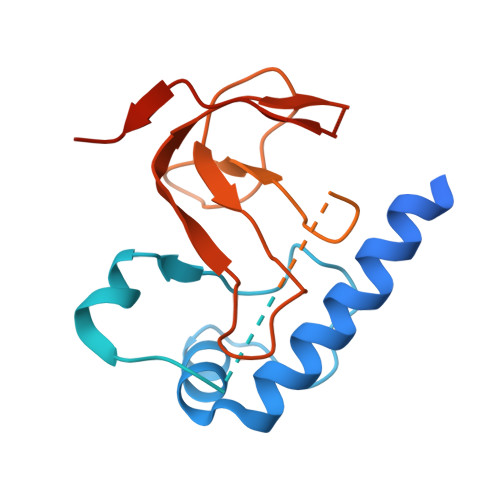
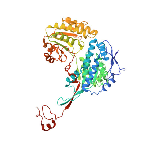
The discovery of reduced flavin mononucleotide and fatty aldehydes as essential factors of light emission facilitated study of bacterial luminescence. Although the molecular mechanisms underlying bacterial luminescence have been studied for more than 60 years, the structure of the bacterial fatty acid reductase complex remains unclear. Here, we report the cryo-EM structure of the Photobacterium phosphoreum fatty acid reductase complex LuxC-LuxE to a resolution of 2.79 Å. We show that the active site Lys238/Arg355 pair of LuxE is >30 Å from the active site Cys296 of LuxC, implying that catalysis relies on a large conformational change. Furthermore, mutagenesis and biochemical experiments support that the L-shaped cleft inside LuxC plays an important role in substrate binding and reaction. We obtained a series of mutants with significantly improved activity as measured by in vitro bioluminescence assays and demonstrated that the double mutant W111A/F483K displayed the highest activity (370% of the WT). Our results indicated that the activity of LuxC significantly affects the bacterial bioluminescence reaction. Finally, we expressed this mutated lux operon in Escherichia coli but observed that the in vivo concentrations of ATP and NADPH limited the enzyme activity; thus, we conclude that the luminous intensity mainly depends on the level of metabolic energy.
- Shenzhen Key Laboratory of Marine Bioresource and Eco-Environmental Science, College of Life Sciences and Oceanography, Shenzhen University, Shenzhen, China; Key Laboratory of Optoelectronic Devices and Systems, College of Physics and Optoelectronic Engineering, Shenzhen University, Shenzhen, China.
Organizational Affiliation: