7X7U | pdb_00007x7u

Cryo-EM structure of SARS-CoV-2 Delta variant spike protein in complex with three nAbs X01, X10 and X17


Experimental Data Snapshot

  • Method: ELECTRON MICROSCOPY
  • Resolution: 3.77 Å
  • Aggregation State: PARTICLE 
  • Reconstruction Method: SINGLE PARTICLE 

wwPDB Validation 3D Report Full Report

Validation slider image for 7X7U

This is version 1.2 of the entry. See complete history

Literature

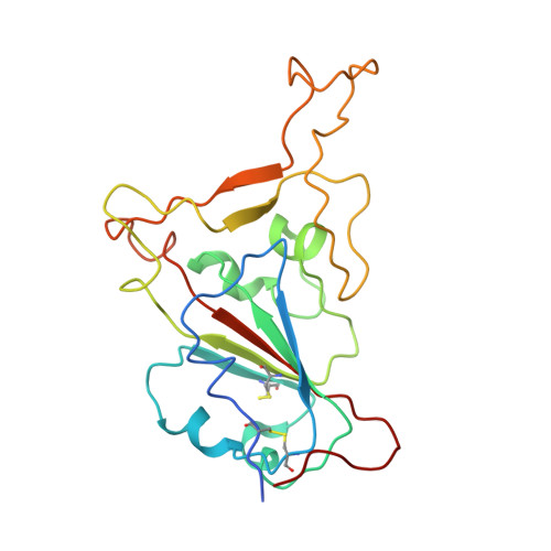
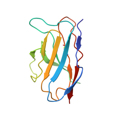
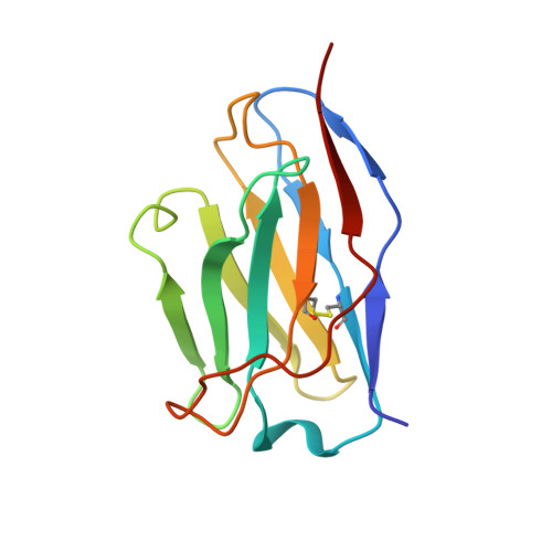
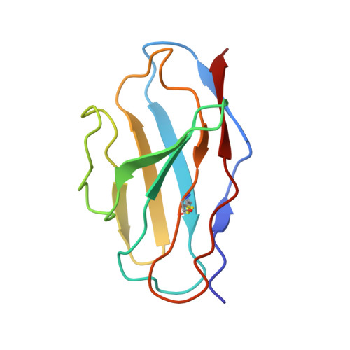
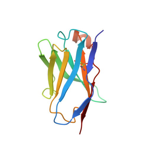
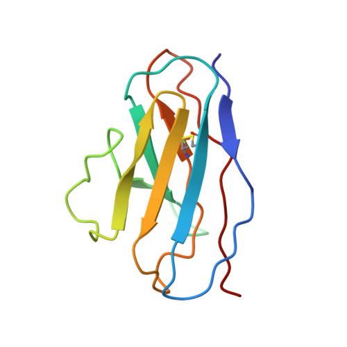
The neutralizing breadth of antibodies targeting diverse conserved epitopes between SARS-CoV and SARS-CoV-2.

Xiong, H.Sun, H.Wang, S.Yuan, L.Liu, L.Zhu, Y.Zhang, J.Huang, Y.Qi, R.Jiang, Y.Ma, J.Zhou, M.Ma, Y.Fu, R.Yan, S.Yue, M.Wu, Y.Wei, M.Wang, Y.Li, T.Wang, Y.Zheng, Z.Yu, H.Cheng, T.Li, S.Yuan, Q.Zhang, J.Guan, Y.Zheng, Q.Zhang, T.Xia, N.

(2022) Proc Natl Acad Sci U S A 119: e2204256119-e2204256119

  • DOI: https://doi.org/10.1073/pnas.2204256119
  • Primary Citation Related Structures: 
    7X7T, 7X7U, 7X7V

  • PubMed Abstract: 

    Antibody therapeutics for the treatment of COVID-19 have been highly successful. However, the recent emergence of the Omicron variant has posed a challenge, as it evades detection by most existing SARS-CoV-2 neutralizing antibodies (nAbs). Here, we successfully generated a panel of SARS-CoV-2/SARS-CoV cross-neutralizing antibodies by sequential immunization of the two pseudoviruses. Of the potential candidates, we found that nAbs X01, X10, and X17 offer broad neutralizing potential against most variants of concern, with X17 further identified as a Class 5 nAb with undiminished neutralization against the Omicron variant. Cryo-electron microscopy structures of the three antibodies together in complex with each of the spike proteins of the prototypical SARS-CoV, SARS-CoV-2, and Delta and Omicron variants of SARS-CoV-2 defined three nonoverlapping conserved epitopes on the receptor-binding domain. The triple-antibody mixture exhibited enhanced resistance to viral evasion and effective protection against infection of the Beta variant in hamsters. Our findings will aid the development of antibody therapeutics and broad vaccines against SARS-CoV-2 and its emerging variants.


  • Organizational Affiliation
    • State Key Laboratory of Molecular Vaccinology and Molecular Diagnostics, National Institute of Diagnostics and Vaccine Development in Infectious Diseases, School of Public Health, School of Life Sciences, Xiamen University, Xiamen 361102, China.

Macromolecule Content 

  • Total Structure Weight: 98.49 kDa 
  • Atom Count: 6,862 
  • Modeled Residue Count: 878 
  • Deposited Residue Count: 888 
  • Unique protein chains: 7

Macromolecules

Find similar proteins by:|  3D Structure
Entity ID: 1
MoleculeChains  Sequence LengthOrganismDetailsImage
Spike protein S1A [auth G]204Severe acute respiratory syndrome coronavirus 2Mutation(s): 2 
Gene Names: S2
UniProt
Find proteins for P0DTC2 (Severe acute respiratory syndrome coronavirus 2)
Explore P0DTC2 
Go to UniProtKB:  P0DTC2
Entity Groups
Sequence Clusters30% Identity50% Identity70% Identity90% Identity95% Identity100% Identity
UniProt GroupP0DTC2
Glycosylation
Glycosylation Sites: 1Go to GlyGen: P0DTC2-1
Sequence Annotations
Expand
Reference Sequence
Find similar proteins by:|  3D Structure
Entity ID: 2
MoleculeChains  Sequence LengthOrganismDetailsImage
X10 light chainB [auth L]111Mus musculusMutation(s): 0 
Find similar proteins by:|  3D Structure
Entity ID: 3
MoleculeChains  Sequence LengthOrganismDetailsImage
X10 heavy chainC [auth H]121Mus musculusMutation(s): 0 
Find similar proteins by:|  3D Structure
Entity ID: 4
MoleculeChains  Sequence LengthOrganismDetailsImage
X17 light chainD [auth B]107Mus musculusMutation(s): 0 
Find similar proteins by:|  3D Structure
Entity ID: 5
MoleculeChains  Sequence LengthOrganismDetailsImage
X17 heavy chainE [auth A]119Mus musculusMutation(s): 0 
Find similar proteins by:|  3D Structure
Entity ID: 6
MoleculeChains  Sequence LengthOrganismDetailsImage
X01 light chainF [auth D]107Mus musculusMutation(s): 0 
Find similar proteins by:|  3D Structure
Entity ID: 7
MoleculeChains  Sequence LengthOrganismDetailsImage
X01 heavy chainG [auth C]119Mus musculusMutation(s): 0 

Small Molecules

Ligands 1 Unique
IDChains Name / Formula / InChI Key2D Diagram3D Interactions
NAG
(Subject of Investigation/LOI)

Query on NAG


Download:Ideal Coordinates CCD File
H [auth G]2-acetamido-2-deoxy-beta-D-glucopyranose
C8 H15 N O6
OVRNDRQMDRJTHS-FMDGEEDCSA-N

Experimental Data & Validation

Experimental Data

  • Method: ELECTRON MICROSCOPY
  • Resolution: 3.77 Å
  • Aggregation State: PARTICLE 
  • Reconstruction Method: SINGLE PARTICLE 

Structure Validation

View Full Validation Report


Entry History 

& Funding Information

Deposition Data


Funding OrganizationLocationGrant Number
Not funded--

Revision History  (Full details and data files)

  • Version 1.0: 2022-08-17
    Type: Initial release
  • Version 1.1: 2022-11-23
    Changes: Database references
  • Version 1.2: 2024-11-06
    Changes: Data collection, Structure summary