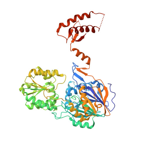
7WNT

RNase J from Mycobacterium tuberculosis


Experimental Data Snapshot

  • Method: X-RAY DIFFRACTION
  • Resolution: 2.44 Å
  • R-Value Free: 0.251 
  • R-Value Work: 0.219 
  • R-Value Observed: 0.221 

wwPDB Validation   3D Report Full Report


This is version 1.1 of the entry. See complete history


Literature

Structural ans functional study of RNase J from Mycobacterium tuberculosis

Li, J.Hu, J.Bao, L.Zhan, B.

To be published.

Macromolecules
Find similar proteins by:  (by identity cutoff)  |  3D Structure
Entity ID: 1
MoleculeChains Sequence LengthOrganismDetailsImage
Ribonuclease J558Mycobacterium tuberculosis H37RvMutation(s): 0 
Gene Names: rnj
EC: 3.1 (PDB Primary Data), 3.5.2.6 (PDB Primary Data)
UniProt
Find proteins for P9WGZ9 (Mycobacterium tuberculosis (strain ATCC 25618 / H37Rv))
Explore P9WGZ9 
Go to UniProtKB:  P9WGZ9
Entity Groups  
Sequence Clusters30% Identity50% Identity70% Identity90% Identity95% Identity100% Identity
UniProt GroupP9WGZ9
Sequence Annotations
Expand
  • Reference Sequence
Experimental Data & Validation

Experimental Data

  • Method: X-RAY DIFFRACTION
  • Resolution: 2.44 Å
  • R-Value Free: 0.251 
  • R-Value Work: 0.219 
  • R-Value Observed: 0.221 
  • Space Group: P 64 2 2
Unit Cell:
Length ( Å )Angle ( ˚ )
a = 128.52α = 90
b = 128.52β = 90
c = 184.276γ = 120
Software Package:
Software NamePurpose
XDSdata reduction
XDSdata scaling
PHENIXphasing
PHENIXrefinement

Structure Validation

View Full Validation Report


Entry History & Funding Information

Deposition Data


Funding OrganizationLocationGrant Number
Ministry of Science and Technology (MoST, China)China2016YFA0500600

Revision History  (Full details and data files)

  • Version 1.0: 2023-01-25
    Type: Initial release
  • Version 1.1: 2023-11-29
    Changes: Data collection, Refinement description