Structural analysis of LpqY, a substrate-binding protein from the SugABC transporter of Mycobacterium tuberculosis, provides insights into its trehalose specificity.
Sharma, D., Singh, M., Kaur, P., Das, U.(2022) Acta Crystallogr D Struct Biol 78: 835-845
- PubMed: 35775983 Search on PubMed
- DOI: https://doi.org/10.1107/S2059798322005290
- Primary Citation Related Structures:
7WCJ, 7WDA - PubMed Abstract:
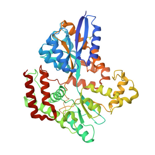
The LpqY-SugABC transporter of Mycobacterium tuberculosis (Mtb) salvages residual trehalose across the cell membrane, which is otherwise lost during the formation of cell-wall glycoconjugates in the periplasm. LpqY, a substrate-binding protein from the SugABC transporter, acts as the primary receptor for the recognition of trehalose, leading to its transport across the cell membrane. Since trehalose is crucial for the survival and virulence of Mtb, trehalose receptors should serve as important targets for novel drug design against tuberculosis. In order to comprehend the detailed architecture and substrate specificity, the first crystal structures of both apo and trehalose-bound forms of M. tuberculosis LpqY (Mtb-LpqY) are presented here at 2.2 and 1.9 Å resolution, respectively. The structure exhibits an N-lobe and C-lobe and is predominantly composed of a globular α/β domain connected by a flexible hinge region concealing a deep binding cleft. Although the trehalose-bound form of Mtb-LpqY revealed an open ligand-bound conformation, the glucose moieties of trehalose are seen to be strongly held in place by direct and water-mediated hydrogen bonds within the binding cavity, producing a K d of 6.58 ± 1.21 µM. These interactions produce a distinct effect on the stereoselectivity for the α-1,1-glycosidic linkage of trehalose. Consistent with the crystal structure, molecular-dynamics simulations further validated Asp43, Asp97 and Asn151 as key residues responsible for strong and stable interactions throughout a 1 µs time frame, thus capturing trehalose in the binding cavity. Collectively, the results provide detailed insights into how the structure and dynamics of Mtb-LpqY enable it to specifically bind trehalose in a relaxed conformation state.
- Department of Biophysics, All India Institute of Medical Sciences, Ansari Nagar, New Delhi 110029, India.
Organizational Affiliation: