Molecular Basis of Mink ACE2 Binding to SARS-CoV-2 and Its Mink-Derived Variants.
Su, C., He, J., Han, P., Bai, B., Li, D., Cao, J., Tian, M., Hu, Y., Zheng, A., Niu, S., Chen, Q., Rong, X., Zhang, Y., Li, W., Qi, J., Zhao, X., Yang, M., Wang, Q., Gao, G.F.(2022) J Virol 96: e0081422-e0081422
- PubMed: 36000849 Search on PubMedSearch on PubMed Central
- DOI: https://doi.org/10.1128/jvi.00814-22
- Primary Citation Related Structures:
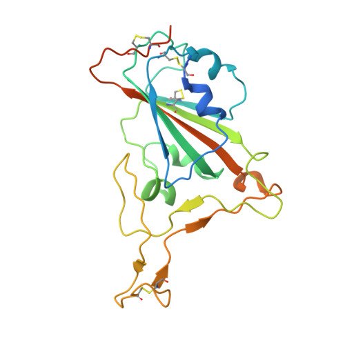
7W8S, 7WA1 - PubMed Abstract:
Severe acute respiratory syndrome coronavirus 2 (SARS-CoV-2) is transmitted between humans and minks, and some mutations in the spike (S) protein, especially in the receptor-binding domain (RBD), have been identified in mink-derived viruses. Here, we examined binding of the mink angiotensin-converting enzyme 2 (ACE2) receptor to mink-derived and important human-originating variants, and we demonstrated that most of the RBD variants increased the binding affinities to mink ACE2 (mkACE2). Cryo-electron microscopy structures of the mkACE2-RBD Y453F (with a Y-to-F change at position 453) and mkACE2-RBD F486L complexes helped identify the key residues that facilitate changes in mkACE2 binding affinity. Additionally, the data indicated that the Y453F and F486L mutations reduced the binding affinities to some human monoclonal antibodies, and human vaccinated sera efficiently prevented infection of human cells by pseudoviruses expressing Y453F, F486L, or N501T RBD. Our findings provide an important molecular mechanism for the rapid adaptation of SARS-CoV-2 in minks and highlight the potential influence of the main mink-originating variants for humans. IMPORTANCE Severe acute respiratory syndrome coronavirus 2 (SARS-CoV-2) has a broad range of hosts. Mink-derived SARS-CoV-2 can transmit back to humans. There is an urgent need to understand the binding mechanism of mink-derived SARS-CoV-2 variants to mink receptor. In this study, we identified all mutations in the receptor-binding domain (RBD) of spike (S) protein from mink-derived SARS-CoV-2, and we demonstrated the enhanced binding affinity of mink angiotensin-converting enzyme 2 (ACE2) to most of the mink-derived RBD variants as well as important human-originating RBD variants. Cryo-electron microscopy structures revealed that the Y453F and F486L mutations enhanced the binding forces in the interaction interface. In addition, Y453F and F486L mutations reduced the binding affinities to some human monoclonal antibodies, and the SARS-CoV-2 pseudoviruses with Y453F, F486L, or N501T mutations were neutralized by human vaccinated sera. Therefore, our results provide valuable information for understanding the cross-species transmission mechanism of SARS-CoV-2.
- Department of Biomedical Sciences, City University of Hong Kong, Hong Kong, China.
Organizational Affiliation: