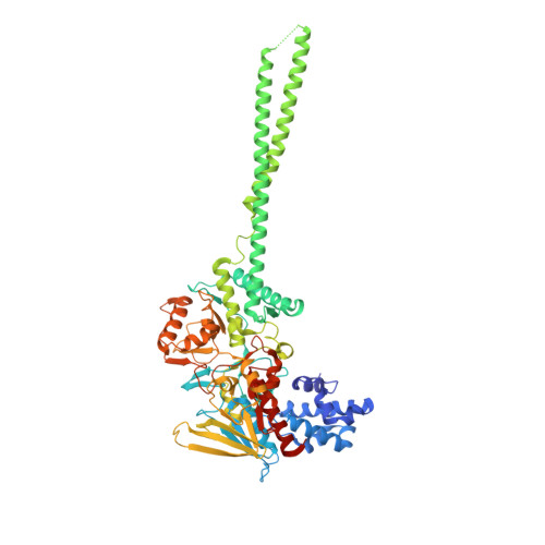
7W3L

Crystal structure of LSD1 in complex with cis-4-Br-2,5-F2-PCPA (S1024)


Experimental Data Snapshot

  • Method: X-RAY DIFFRACTION
  • Resolution: 2.51 Å
  • R-Value Free: 0.216 
  • R-Value Work: 0.178 
  • R-Value Observed: 0.181 

wwPDB Validation   3D Report Full Report


Ligand Structure Quality Assessment 


This is version 1.2 of the entry. See complete history


Literature

Structure-Activity Relationship and In Silico Evaluation of cis- and trans-PCPA-Derived Inhibitors of LSD1 and LSD2

Niwa, H.Watanabe, C.Sato, S.Harada, T.Watanabe, H.Tabusa, R.Fukasawa, S.Shiobara, A.Hashimoto, T.Ohno, O.Nakamura, K.Tsuganezawa, K.Tanaka, A.Shirouzu, M.Honma, T.Matsuno, K.Umehara, T.

(2022) ACS Med Chem Lett 13: 1485-1492


Macromolecules
Find similar proteins by:  (by identity cutoff)  |  3D Structure
Entity ID: 1
MoleculeChains Sequence LengthOrganismDetailsImage
Lysine-specific histone demethylase 1A669Homo sapiensMutation(s): 0 
Gene Names: KDM1AAOF2KDM1KIAA0601LSD1
EC: 1.14.99.66
UniProt & NIH Common Fund Data Resources
Find proteins for O60341 (Homo sapiens)
Explore O60341 
Go to UniProtKB:  O60341
PHAROS:  O60341
GTEx:  ENSG00000004487 
Entity Groups  
Sequence Clusters30% Identity50% Identity70% Identity90% Identity95% Identity100% Identity
UniProt GroupO60341
Sequence Annotations
Expand
  • Reference Sequence
Small Molecules
Ligands 4 Unique
IDChains Name / Formula / InChI Key2D Diagram3D Interactions
FAD
Query on FAD

Download Ideal Coordinates CCD File 
B [auth A]FLAVIN-ADENINE DINUCLEOTIDE
C27 H33 N9 O15 P2
VWWQXMAJTJZDQX-UYBVJOGSSA-N
8A2
Query on 8A2

Download Ideal Coordinates CCD File 
C [auth A]3-[4-bromanyl-2,5-bis(fluoranyl)phenyl]propanal
C9 H7 Br F2 O
JXYLPBJZJRAAJO-UHFFFAOYSA-N
TLA
Query on TLA

Download Ideal Coordinates CCD File 
D [auth A]L(+)-TARTARIC ACID
C4 H6 O6
FEWJPZIEWOKRBE-JCYAYHJZSA-N
GOL
Query on GOL

Download Ideal Coordinates CCD File 
E [auth A]
F [auth A]
G [auth A]
H [auth A]
I [auth A]
E [auth A],
F [auth A],
G [auth A],
H [auth A],
I [auth A],
J [auth A]
GLYCEROL
C3 H8 O3
PEDCQBHIVMGVHV-UHFFFAOYSA-N
Experimental Data & Validation

Experimental Data

  • Method: X-RAY DIFFRACTION
  • Resolution: 2.51 Å
  • R-Value Free: 0.216 
  • R-Value Work: 0.178 
  • R-Value Observed: 0.181 
  • Space Group: P 61 2 2
Unit Cell:
Length ( Å )Angle ( ˚ )
a = 186.433α = 90
b = 186.433β = 90
c = 107.457γ = 120
Software Package:
Software NamePurpose
PHENIXrefinement
XDSdata reduction
Aimlessdata scaling
PHENIXphasing

Structure Validation

View Full Validation Report


Ligand Structure Quality Assessment 


Entry History & Funding Information

Deposition Data


Funding OrganizationLocationGrant Number
Japan Society for the Promotion of Science (JSPS)Japan21K06461, 20H03388, 20K21406

Revision History  (Full details and data files)

  • Version 1.0: 2022-09-14
    Type: Initial release
  • Version 1.1: 2022-09-21
    Changes: Database references, Structure summary
  • Version 1.2: 2023-11-29
    Changes: Data collection, Refinement description