7VSG

Cryo-EM structure of a human ATP11C-CDC50A flippase reconstituted in the Nanodisc in PtdSer-occluded E2-Pi state.


Experimental Data Snapshot

  • Method: ELECTRON MICROSCOPY
  • Resolution: 3.90 Å
  • Aggregation State: PARTICLE 
  • Reconstruction Method: SINGLE PARTICLE 

wwPDB Validation   3D Report Full Report


This is version 1.1 of the entry. See complete history


Literature

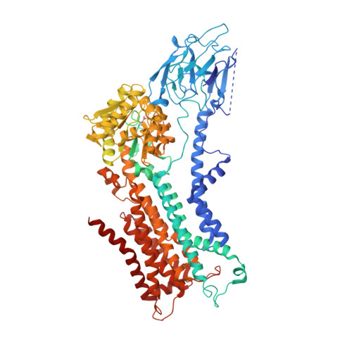
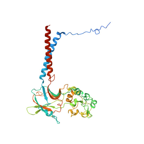
Cryo-EM of the ATP11C flippase reconstituted in Nanodiscs shows a distended phospholipid bilayer inner membrane around transmembrane helix 2.

Nakanishi, H.Hayashida, K.Nishizawa, T.Oshima, A.Abe, K.

(2022) J Biol Chem 298: 101498-101498

  • DOI: https://doi.org/10.1016/j.jbc.2021.101498
  • Primary Citation of Related Structures:  
    7VSG, 7VSH

  • PubMed Abstract: 

    ATP11C is a member of the P4-ATPase flippase family that mediates translocation of phosphatidylserine (PtdSer) across the lipid bilayer. In order to characterize the structure and function of ATP11C in a model natural lipid environment, we revisited and optimized a quick procedure for reconstituting ATP11C into Nanodiscs using methyl-β-cyclodextrin as a reagent for the detergent removal. ATP11C was efficiently reconstituted with the endogenous lipid, or the mixture of endogenous lipid and synthetic dioleoylphosphatidylcholine (DOPC)/dioleoylphosphatidylserine (DOPS), all of which retained the ATPase activity. We obtained 3.4 Å and 3.9 Å structures using single-particle cryo-electron microscopy (cryo-EM) of AlF- and BeF-stabilized ATP11C transport intermediates, respectively, in a bilayer containing DOPS. We show that the latter exhibited a distended inner membrane around ATP11C transmembrane helix 2, possibly reflecting the perturbation needed for phospholipid release to the lipid bilayer. Our structures of ATP11C in the lipid membrane indicate that the membrane boundary varies upon conformational changes of the enzyme and is no longer flat around the protein, a change that likely contributes to phospholipid translocation across the membrane leaflets.


  • Organizational Affiliation

    Cellular and Structural Physiology Institute, Nagoya University, Nagoya, Japan.


Macromolecules
Find similar proteins by:  (by identity cutoff)  |  3D Structure
Entity ID: 1
MoleculeChains Sequence LengthOrganismDetailsImage
Phospholipid-transporting ATPase IG1,084Homo sapiensMutation(s): 1 
Gene Names: ATP11CATPIGATPIQ
EC: 7.6.2.1
Membrane Entity: Yes 
UniProt & NIH Common Fund Data Resources
Find proteins for Q8NB49 (Homo sapiens)
Explore Q8NB49 
Go to UniProtKB:  Q8NB49
PHAROS:  Q8NB49
GTEx:  ENSG00000101974 
Entity Groups  
Sequence Clusters30% Identity50% Identity70% Identity90% Identity95% Identity100% Identity
UniProt GroupQ8NB49
Sequence Annotations
Expand
  • Reference Sequence
Find similar proteins by:  (by identity cutoff)  |  3D Structure
Entity ID: 2
MoleculeChains Sequence LengthOrganismDetailsImage
Cell cycle control protein 50A361Homo sapiensMutation(s): 0 
Gene Names: CDC50A
Membrane Entity: Yes 
UniProt & NIH Common Fund Data Resources
Find proteins for Q9NV96 (Homo sapiens)
Explore Q9NV96 
Go to UniProtKB:  Q9NV96
PHAROS:  Q9NV96
GTEx:  ENSG00000112697 
Entity Groups  
Sequence Clusters30% Identity50% Identity70% Identity90% Identity95% Identity100% Identity
UniProt GroupQ9NV96
Sequence Annotations
Expand
  • Reference Sequence
Oligosaccharides

Help

Entity ID: 3
MoleculeChains Length2D Diagram Glycosylation3D Interactions
alpha-D-mannopyranose-(1-4)-2-acetamido-2-deoxy-beta-D-glucopyranose-(1-4)-2-acetamido-2-deoxy-beta-D-glucopyranoseC [auth D]3N-Glycosylation
Glycosylation Resources
GlyTouCan:  G62182OO
GlyCosmos:  G62182OO
GlyGen:  G62182OO
Small Molecules
Ligands 2 Unique
IDChains Name / Formula / InChI Key2D Diagram3D Interactions
17F (Subject of Investigation/LOI)
Query on 17F

Download Ideal Coordinates CCD File 
D [auth A]O-[(S)-({(2R)-2,3-bis[(9Z)-octadec-9-enoyloxy]propyl}oxy)(hydroxy)phosphoryl]-L-serine
C42 H78 N O10 P
WTBFLCSPLLEDEM-JIDRGYQWSA-N
NAG
Query on NAG

Download Ideal Coordinates CCD File 
E [auth B],
F [auth B]
2-acetamido-2-deoxy-beta-D-glucopyranose
C8 H15 N O6
OVRNDRQMDRJTHS-FMDGEEDCSA-N
Modified Residues  1 Unique
IDChains TypeFormula2D DiagramParent
BFD
Query on BFD
A
L-PEPTIDE LINKINGC4 H6 Be F3 N O4ASP
Experimental Data & Validation

Experimental Data

  • Method: ELECTRON MICROSCOPY
  • Resolution: 3.90 Å
  • Aggregation State: PARTICLE 
  • Reconstruction Method: SINGLE PARTICLE 

Structure Validation

View Full Validation Report


Entry History & Funding Information

Deposition Data


Funding OrganizationLocationGrant Number
Japan Society for the Promotion of Science (JSPS)Japan21K06064
Japan Society for the Promotion of Science (JSPS)Japan21H02426

Revision History  (Full details and data files)

  • Version 1.0: 2021-12-29
    Type: Initial release
  • Version 1.1: 2022-02-16
    Changes: Database references