7VMP

Structure of recombinant RyR2 (Ca2+ dataset, class 2, open state)


Experimental Data Snapshot

  • Method: ELECTRON MICROSCOPY
  • Resolution: 3.50 Å
  • Aggregation State: PARTICLE 
  • Reconstruction Method: SINGLE PARTICLE 

wwPDB Validation   3D Report Full Report


This is version 1.0 of the entry. See complete history


Literature

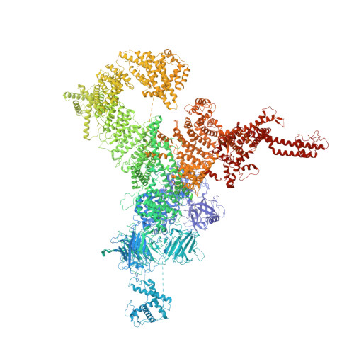
Molecular basis for gating of cardiac ryanodine receptor explains the mechanisms for gain- and loss-of function mutations.

Kobayashi, T.Tsutsumi, A.Kurebayashi, N.Saito, K.Kodama, M.Sakurai, T.Kikkawa, M.Murayama, T.Ogawa, H.

(2022) Nat Commun 13: 2821-2821

  • DOI: https://doi.org/10.1038/s41467-022-30429-x
  • Primary Citation of Related Structures:  
    7VML, 7VMM, 7VMN, 7VMO, 7VMP, 7VMR

  • PubMed Abstract: 

    Cardiac ryanodine receptor (RyR2) is a large Ca 2+ release channel in the sarcoplasmic reticulum and indispensable for excitation-contraction coupling in the heart. RyR2 is activated by Ca 2+ and RyR2 mutations are implicated in severe arrhythmogenic diseases. Yet, the structural basis underlying channel opening and how mutations affect the channel remains unknown. Here, we address the gating mechanism of RyR2 by combining high-resolution structures determined by cryo-electron microscopy with quantitative functional analysis of channels carrying various mutations in specific residues. We demonstrated two fundamental mechanisms for channel gating: interactions close to the channel pore stabilize the channel to prevent hyperactivity and a series of interactions in the surrounding regions is necessary for channel opening upon Ca 2+ binding. Mutations at the residues involved in the former and the latter mechanisms cause gain-of-function and loss-of-function, respectively. Our results reveal gating mechanisms of the RyR2 channel and alterations by pathogenic mutations at the atomic level.


  • Organizational Affiliation

    Department of Cellular and Molecular Pharmacology, Juntendo University Graduate School of Medicine, Tokyo, Japan.


Macromolecules
Find similar proteins by:  (by identity cutoff)  |  3D Structure
Entity ID: 1
MoleculeChains Sequence LengthOrganismDetailsImage
Ryanodine receptor 2A,
C [auth B],
E [auth C],
G [auth D]
4,966Mus musculusMutation(s): 0 
Gene Names: Ryr2
Membrane Entity: Yes 
UniProt & NIH Common Fund Data Resources
Find proteins for E9Q401 (Mus musculus)
Explore E9Q401 
Go to UniProtKB:  E9Q401
IMPC:  MGI:99685
Entity Groups  
Sequence Clusters30% Identity50% Identity70% Identity90% Identity95% Identity100% Identity
UniProt GroupE9Q401
Sequence Annotations
Expand
  • Reference Sequence
Find similar proteins by:  (by identity cutoff)  |  3D Structure
Entity ID: 2
MoleculeChains Sequence LengthOrganismDetailsImage
Peptidyl-prolyl cis-trans isomerase FKBP1BB [auth G],
D [auth H],
F [auth I],
H [auth J]
176Homo sapiensMutation(s): 0 
Gene Names: FKBP1B
EC: 5.2.1.8
UniProt & NIH Common Fund Data Resources
Find proteins for P68106 (Homo sapiens)
Explore P68106 
Go to UniProtKB:  P68106
PHAROS:  P68106
GTEx:  ENSG00000119782 
Entity Groups  
Sequence Clusters30% Identity50% Identity70% Identity90% Identity95% Identity100% Identity
UniProt GroupP68106
Sequence Annotations
Expand
  • Reference Sequence
Experimental Data & Validation

Experimental Data

  • Method: ELECTRON MICROSCOPY
  • Resolution: 3.50 Å
  • Aggregation State: PARTICLE 
  • Reconstruction Method: SINGLE PARTICLE 

Structure Validation

View Full Validation Report


Entry History & Funding Information

Deposition Data


Funding OrganizationLocationGrant Number
Japan Society for the Promotion of Science (JSPS)JapanJP16H04748
Japan Society for the Promotion of Science (JSPS)JapanJP19K07105
Japan Society for the Promotion of Science (JSPS)Japan19H03404
Japan Society for the Promotion of Science (JSPS)JapanJP21H02411
Japan Agency for Medical Research and Development (AMED)JapanJP21am0101080
Japan Agency for Medical Research and Development (AMED)Japan19ek0109202

Revision History  (Full details and data files)

  • Version 1.0: 2022-08-10
    Type: Initial release