7VGG

Cryo-EM structure of Ultraviolet-B activated UVR8 in complex with COP1


Experimental Data Snapshot

  • Method: ELECTRON MICROSCOPY
  • Resolution: 3.10 Å
  • Aggregation State: PARTICLE 
  • Reconstruction Method: SINGLE PARTICLE 

wwPDB Validation   3D Report Full Report


This is version 1.1 of the entry. See complete history


Literature

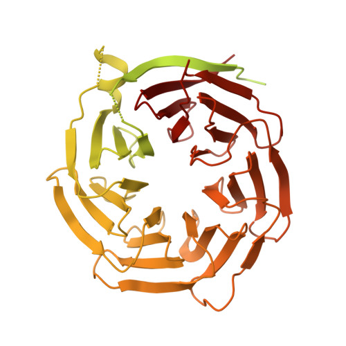
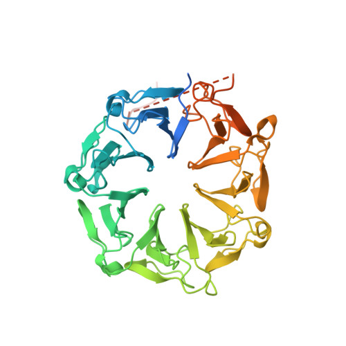
Structural insight into UV-B-activated UVR8 bound to COP1.

Wang, Y.Wang, L.Guan, Z.Chang, H.Ma, L.Shen, C.Qiu, L.Yan, J.Zhang, D.Li, J.Deng, X.W.Yin, P.

(2022) Sci Adv 8: eabn3337-eabn3337

  • DOI: https://doi.org/10.1126/sciadv.abn3337
  • Primary Citation of Related Structures:  
    7VGG

  • PubMed Abstract: 

    The CONSTITUTIVE PHOTOMORPHOGENIC 1-SUPPRESSOR OF PHYA-105 (COP1-SPA) complex is a central repressor of photomorphogenesis. This complex acts as an E3 ubiquitin ligase downstream of various light signaling transduced from multiple photoreceptors in plants. How the COP1-SPA activity is regulated by divergent light-signaling pathways remains largely elusive. Here, we reproduced the regulation pathway of COP1-SPA in ultraviolet-B (UV-B) signaling in vitro and determined the cryo-electron microscopy structure of UV-B receptor UVR8 in complex with COP1. The complex formation is mediated by two-interface interactions between UV-B-activated UVR8 and COP1. Both interfaces are essential for the competitive binding of UVR8 against the signaling hub component HY5 to the COP1-SPA complex. We also show that RUP2 dissociates UVR8 from the COP1-SPA4 1-464 -UVR8 complex and facilitates its redimerization. Our results support a UV-B signaling model that the COP1-SPA activity is repressed by UV-B-activated UVR8 and derepressed by RUP2, owing to competitive binding, and provide a framework for studying the regulatory roles of distinct photoreceptors on photomorphogenesis.


  • Organizational Affiliation

    National Key Laboratory of Crop Genetic Improvement, Hubei Hongshan Laboratory, Huazhong Agricultural University, Wuhan 430070, China.


Macromolecules
Find similar proteins by:  (by identity cutoff)  |  3D Structure
Entity ID: 1
MoleculeChains Sequence LengthOrganismDetailsImage
E3 ubiquitin-protein ligase COP1704Arabidopsis thalianaMutation(s): 0 
Gene Names: COP1
EC: 2.3.2.27
UniProt
Find proteins for P43254 (Arabidopsis thaliana)
Explore P43254 
Go to UniProtKB:  P43254
Entity Groups  
Sequence Clusters30% Identity50% Identity70% Identity90% Identity95% Identity100% Identity
UniProt GroupP43254
Sequence Annotations
Expand
  • Reference Sequence
Find similar proteins by:  (by identity cutoff)  |  3D Structure
Entity ID: 2
MoleculeChains Sequence LengthOrganismDetailsImage
Ultraviolet-B receptor UVR8468Arabidopsis thalianaMutation(s): 0 
Gene Names: UVR8
UniProt
Find proteins for Q9FN03 (Arabidopsis thaliana)
Explore Q9FN03 
Go to UniProtKB:  Q9FN03
Entity Groups  
Sequence Clusters30% Identity50% Identity70% Identity90% Identity95% Identity100% Identity
UniProt GroupQ9FN03
Sequence Annotations
Expand
  • Reference Sequence
Experimental Data & Validation

Experimental Data

  • Method: ELECTRON MICROSCOPY
  • Resolution: 3.10 Å
  • Aggregation State: PARTICLE 
  • Reconstruction Method: SINGLE PARTICLE 

Structure Validation

View Full Validation Report


Entry History & Funding Information

Deposition Data


Funding OrganizationLocationGrant Number
National Natural Science Foundation of China (NSFC)China2018YFA0507700

Revision History  (Full details and data files)

  • Version 1.0: 2022-05-04
    Type: Initial release
  • Version 1.1: 2022-05-11
    Changes: Database references