7VGG | pdb_00007vgg

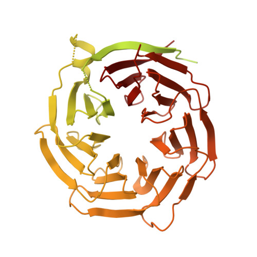
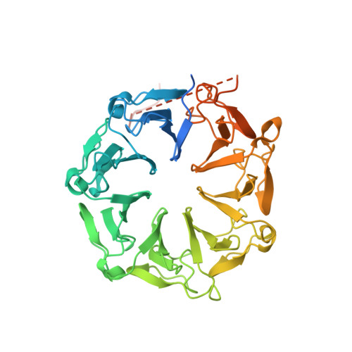
Cryo-EM structure of Ultraviolet-B activated UVR8 in complex with COP1


Experimental Data Snapshot

  • Method: ELECTRON MICROSCOPY
  • Resolution: 3.10 Å
  • Aggregation State: PARTICLE 
  • Reconstruction Method: SINGLE PARTICLE 

wwPDB Validation 3D Report Full Report

Validation slider image for 7VGG

This is version 1.3 of the entry. See complete history. 

Literature

Structural insight into UV-B-activated UVR8 bound to COP1.

Wang, Y., Wang, L., Guan, Z., Chang, H., Ma, L., Shen, C., Qiu, L., Yan, J., Zhang, D., Li, J., Deng, X.W., Yin, P.

(2022) Sci Adv 8: eabn3337-eabn3337

  • DOI: https://doi.org/10.1126/sciadv.abn3337
  • Primary Citation Related Structures: 
    7VGG

  • PubMed Abstract: 

    The CONSTITUTIVE PHOTOMORPHOGENIC 1-SUPPRESSOR OF PHYA-105 (COP1-SPA) complex is a central repressor of photomorphogenesis. This complex acts as an E3 ubiquitin ligase downstream of various light signaling transduced from multiple photoreceptors in plants. How the COP1-SPA activity is regulated by divergent light-signaling pathways remains largely elusive. Here, we reproduced the regulation pathway of COP1-SPA in ultraviolet-B (UV-B) signaling in vitro and determined the cryo-electron microscopy structure of UV-B receptor UVR8 in complex with COP1. The complex formation is mediated by two-interface interactions between UV-B-activated UVR8 and COP1. Both interfaces are essential for the competitive binding of UVR8 against the signaling hub component HY5 to the COP1-SPA complex. We also show that RUP2 dissociates UVR8 from the COP1-SPA4 1-464 -UVR8 complex and facilitates its redimerization. Our results support a UV-B signaling model that the COP1-SPA activity is repressed by UV-B-activated UVR8 and derepressed by RUP2, owing to competitive binding, and provide a framework for studying the regulatory roles of distinct photoreceptors on photomorphogenesis.


  • Organizational Affiliation: 
    • National Key Laboratory of Crop Genetic Improvement, Hubei Hongshan Laboratory, Huazhong Agricultural University, Wuhan 430070, China.

Macromolecule Content 

  • Total Structure Weight: 129.96 kDa 
  • Atom Count: 5,274 
  • Modeled Residue Count: 680 
  • Deposited Residue Count: 1,172 
  • Unique protein chains: 2

Macromolecules

Find similar proteins by:|  3D Structure
Entity ID: 1
MoleculeChains  Sequence LengthOrganismDetailsImage
E3 ubiquitin-protein ligase COP1704Arabidopsis thalianaMutation(s): 0 
Gene Names: COP1
EC: 2.3.2.27
UniProt
Find proteins for P43254 (Arabidopsis thaliana)
Explore P43254 
Go to UniProtKB:  P43254
Entity Groups
Sequence Clusters30% Identity50% Identity70% Identity90% Identity95% Identity100% Identity
UniProt GroupP43254
Sequence Annotations
Expand
Reference Sequence
Find similar proteins by:|  3D Structure
Entity ID: 2
MoleculeChains  Sequence LengthOrganismDetailsImage
Ultraviolet-B receptor UVR8468Arabidopsis thalianaMutation(s): 0 
Gene Names: UVR8
UniProt
Find proteins for Q9FN03 (Arabidopsis thaliana)
Explore Q9FN03 
Go to UniProtKB:  Q9FN03
Entity Groups
Sequence Clusters30% Identity50% Identity70% Identity90% Identity95% Identity100% Identity
UniProt GroupQ9FN03
Sequence Annotations
Expand
Reference Sequence

Experimental Data & Validation

Experimental Data

  • Method: ELECTRON MICROSCOPY
  • Resolution: 3.10 Å
  • Aggregation State: PARTICLE 
  • Reconstruction Method: SINGLE PARTICLE 
EM Software:
TaskSoftware PackageVersion
MODEL REFINEMENTPHENIX

Structure Validation

View Full Validation Report


Entry History 

& Funding Information

Deposition Data


Funding OrganizationLocationGrant Number
National Natural Science Foundation of China (NSFC)China2018YFA0507700

Revision History  (Full details and data files)

  • Version 1.0: 2022-05-04
    Type: Initial release
  • Version 1.1: 2022-05-11
    Changes: Database references
  • Version 1.2: 2024-06-19
    Changes: Data collection
  • Version 1.3: 2025-06-18
    Changes: Data collection, Structure summary