7USY

Structure of C. elegans TMC-1 complex with ARRD-6


Experimental Data Snapshot

  • Method: ELECTRON MICROSCOPY
  • Resolution: 3.54 Å
  • Resolution: 3.54 Å
  • Resolution: 3.54 Å
  • Resolution: 3.54 Å
  • Aggregation State: PARTICLE 
  • Reconstruction Method: SINGLE PARTICLE 

wwPDB Validation   3D Report Full Report


This is version 3.0 of the entry. See complete history


Literature

Structures of the TMC-1 complex illuminate mechanosensory transduction.

Jeong, H.Clark, S.Goehring, A.Dehghani-Ghahnaviyeh, S.Rasouli, A.Tajkhorshid, E.Gouaux, E.

(2022) Nature 610: 796-803

  • DOI: https://doi.org/10.1038/s41586-022-05314-8
  • Primary Citation of Related Structures:  
    7USW, 7USX, 7USY

  • PubMed Abstract: 

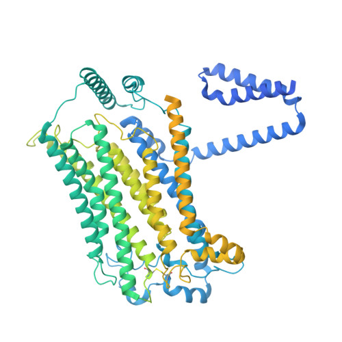
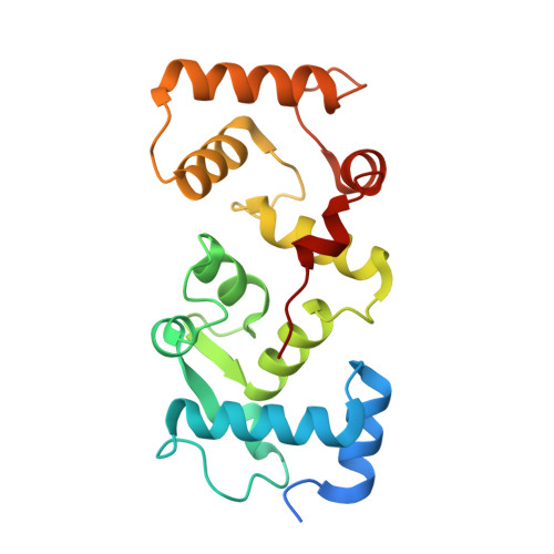
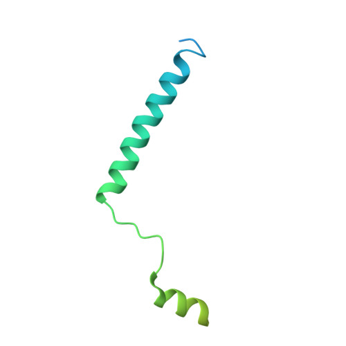
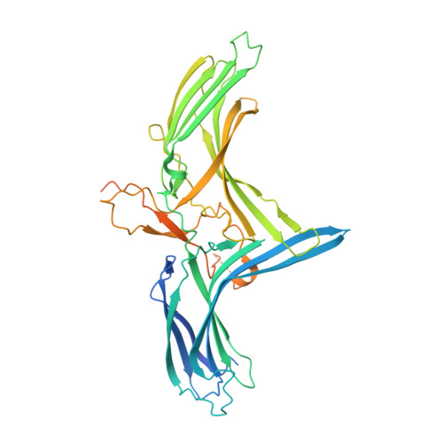
    The initial step in the sensory transduction pathway underpinning hearing and balance in mammals involves the conversion of force into the gating of a mechanosensory transduction channel 1 . Despite the profound socioeconomic impacts of hearing disorders and the fundamental biological significance of understanding mechanosensory transduction, the composition, structure and mechanism of the mechanosensory transduction complex have remained poorly characterized. Here we report the single-particle cryo-electron microscopy structure of the native transmembrane channel-like protein 1 (TMC-1) mechanosensory transduction complex isolated from Caenorhabditis elegans. The two-fold symmetric complex is composed of two copies each of the pore-forming TMC-1 subunit, the calcium-binding protein CALM-1 and the transmembrane inner ear protein TMIE. CALM-1 makes extensive contacts with the cytoplasmic face of the TMC-1 subunits, whereas the single-pass TMIE subunits reside on the periphery of the complex, poised like the handles of an accordion. A subset of complexes additionally includes a single arrestin-like protein, arrestin domain protein (ARRD-6), bound to a CALM-1 subunit. Single-particle reconstructions and molecular dynamics simulations show how the mechanosensory transduction complex deforms the membrane bilayer and suggest crucial roles for lipid-protein interactions in the mechanism by which mechanical force is transduced to ion channel gating.


  • Organizational Affiliation

    Vollum Institute, Oregon Health and Science University, Portland, OR, USA.


Macromolecules
Find similar proteins by:  (by identity cutoff)  |  3D Structure
Entity ID: 1
MoleculeChains Sequence LengthOrganismDetailsImage
Transmembrane channel-like protein 1
A, B
1,285Caenorhabditis elegansMutation(s): 0 
Membrane Entity: Yes 
UniProt
Find proteins for D3KZG3 (Caenorhabditis elegans)
Explore D3KZG3 
Go to UniProtKB:  D3KZG3
Entity Groups  
Sequence Clusters30% Identity50% Identity70% Identity90% Identity95% Identity100% Identity
UniProt GroupD3KZG3
Sequence Annotations
Expand
  • Reference Sequence
Find similar proteins by:  (by identity cutoff)  |  3D Structure
Entity ID: 2
MoleculeChains Sequence LengthOrganismDetailsImage
CALMyrin (Calcium and Integrin Binding protein) homolog
C, E
201Caenorhabditis elegansMutation(s): 0 
UniProt
Find proteins for Q93640 (Caenorhabditis elegans)
Explore Q93640 
Go to UniProtKB:  Q93640
Entity Groups  
Sequence Clusters30% Identity50% Identity70% Identity90% Identity95% Identity100% Identity
UniProt GroupQ93640
Sequence Annotations
Expand
  • Reference Sequence
Find similar proteins by:  (by identity cutoff)  |  3D Structure
Entity ID: 3
MoleculeChains Sequence LengthOrganismDetailsImage
Transmembrane inner ear expressed protein
D, F
117Caenorhabditis elegansMutation(s): 0 
Membrane Entity: Yes 
UniProt
Find proteins for Q9XXE7 (Caenorhabditis elegans)
Explore Q9XXE7 
Go to UniProtKB:  Q9XXE7
Entity Groups  
Sequence Clusters30% Identity50% Identity70% Identity90% Identity95% Identity100% Identity
UniProt GroupQ9XXE7
Sequence Annotations
Expand
  • Reference Sequence
Find similar proteins by:  (by identity cutoff)  |  3D Structure
Entity ID: 4
MoleculeChains Sequence LengthOrganismDetailsImage
ARRestin Domain proteinG [auth J]469Caenorhabditis elegansMutation(s): 0 
UniProt
Find proteins for D1MN74 (Caenorhabditis elegans)
Explore D1MN74 
Go to UniProtKB:  D1MN74
Entity Groups  
Sequence Clusters30% Identity50% Identity70% Identity90% Identity95% Identity100% Identity
UniProt GroupD1MN74
Sequence Annotations
Expand
  • Reference Sequence
Small Molecules
Ligands 5 Unique
IDChains Name / Formula / InChI Key2D Diagram3D Interactions
3PE (Subject of Investigation/LOI)
Query on 3PE

Download Ideal Coordinates CCD File 
K [auth A],
P [auth B]
1,2-Distearoyl-sn-glycerophosphoethanolamine
C41 H82 N O8 P
LVNGJLRDBYCPGB-LDLOPFEMSA-N
PEE (Subject of Investigation/LOI)
Query on PEE

Download Ideal Coordinates CCD File 
J [auth A],
O [auth B]
1,2-dioleoyl-sn-glycero-3-phosphoethanolamine
C41 H78 N O8 P
MWRBNPKJOOWZPW-NYVOMTAGSA-N
PLM (Subject of Investigation/LOI)
Query on PLM

Download Ideal Coordinates CCD File 
T [auth D],
U [auth D],
X [auth F],
Y [auth F]
PALMITIC ACID
C16 H32 O2
IPCSVZSSVZVIGE-UHFFFAOYSA-N
NAG (Subject of Investigation/LOI)
Query on NAG

Download Ideal Coordinates CCD File 
L [auth A],
Q [auth B]
2-acetamido-2-deoxy-beta-D-glucopyranose
C8 H15 N O6
OVRNDRQMDRJTHS-FMDGEEDCSA-N
CA (Subject of Investigation/LOI)
Query on CA

Download Ideal Coordinates CCD File 
H [auth A]
I [auth A]
M [auth B]
N [auth B]
R [auth C]
H [auth A],
I [auth A],
M [auth B],
N [auth B],
R [auth C],
S [auth C],
V [auth E],
W [auth E]
CALCIUM ION
Ca
BHPQYMZQTOCNFJ-UHFFFAOYSA-N
Experimental Data & Validation

Experimental Data

  • Method: ELECTRON MICROSCOPY
  • Resolution: 3.54 Å
  • Resolution: 3.54 Å
  • Resolution: 3.54 Å
  • Resolution: 3.54 Å
  • Aggregation State: PARTICLE 
  • Reconstruction Method: SINGLE PARTICLE 

Structure Validation

View Full Validation Report


Entry History & Funding Information

Deposition Data


Funding OrganizationLocationGrant Number
Howard Hughes Medical Institute (HHMI)United States--

Revision History  (Full details and data files)

  • Version 1.0: 2022-10-19
    Type: Initial release
  • Version 2.0: 2022-10-26
    Changes: Advisory, Atomic model, Author supporting evidence, Data collection, Database references, Derived calculations, Non-polymer description, Structure summary
  • Version 2.1: 2022-11-09
    Changes: Database references
  • Version 3.0: 2023-08-09
    Type: Coordinate replacement
    Reason: Model completeness
    Changes: Advisory, Atomic model, Author supporting evidence, Data collection, Data processing, Derived calculations, Refinement description, Structure summary