7US1

Structure of parkin (R0RB) bound to two phospho-ubiquitin molecules


Experimental Data Snapshot

  • Method: X-RAY DIFFRACTION
  • Resolution: 2.48 Å
  • R-Value Free: 0.258 
  • R-Value Work: 0.229 
  • R-Value Observed: 0.231 

Starting Model: experimental
View more details

wwPDB Validation   3D Report Full Report


This is version 1.2 of the entry. See complete history


Literature

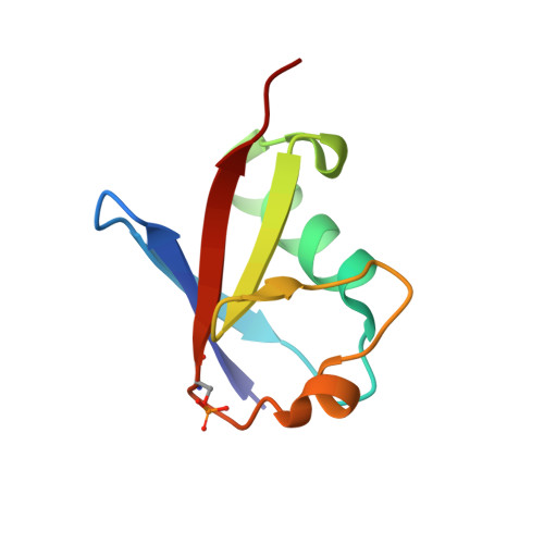
Structure of the second phosphoubiquitin-binding site in parkin.

Fakih, R.Sauve, V.Gehring, K.

(2022) J Biol Chem 298: 102114-102114

  • DOI: https://doi.org/10.1016/j.jbc.2022.102114
  • Primary Citation of Related Structures:  
    7US1

  • PubMed Abstract: 

    Parkin and PINK1 regulate a mitochondrial quality control system that is mutated in some early onset forms of Parkinson's disease. Parkin is an E3 ubiquitin ligase and regulated by the mitochondrial kinase PINK1 via a two-step cascade. PINK1 first phosphorylates ubiquitin, which binds a recruitment site on parkin to localize parkin to damaged mitochondria. In the second step, PINK1 phosphorylates parkin on its ubiquitin-like domain (Ubl), which binds a regulatory site to release ubiquitin ligase activity. Recently, an alternative feed-forward mechanism was identified that bypasses the need for parkin phosphorylation through the binding of a second phosphoubiquitin (pUb) molecule. Here, we report the structure of parkin activated through this feed-forward mechanism. The crystal structure of parkin with pUb bound to both the recruitment and regulatory sites reveals the molecular basis for differences in specificity and affinity of the two sites. We use isothermal titration calorimetry measurements to reveal cooperativity between the two binding sites and the role of linker residues for pUbl binding to the regulatory site. The observation of flexibility in the process of parkin activation offers hope for the future design of small molecules for the treatment of Parkinson's disease.


  • Organizational Affiliation

    Department of Biochemistry and Centre de recherche en biologie structurale, McGill University, Montreal, Quebec, Canada.


Macromolecules
Find similar proteins by:  (by identity cutoff)  |  3D Structure
Entity ID: 1
MoleculeChains Sequence LengthOrganismDetailsImage
E3 ubiquitin-protein ligase parkin240Rattus norvegicusMutation(s): 0 
Gene Names: Park2
EC: 2.3.2.31
UniProt
Find proteins for Q9JK66 (Rattus norvegicus)
Explore Q9JK66 
Go to UniProtKB:  Q9JK66
Entity Groups  
Sequence Clusters30% Identity50% Identity70% Identity90% Identity95% Identity100% Identity
UniProt GroupQ9JK66
Sequence Annotations
Expand
  • Reference Sequence
Find similar proteins by:  (by identity cutoff)  |  3D Structure
Entity ID: 2
MoleculeChains Sequence LengthOrganismDetailsImage
Ubiquitin-G76 deletion
B, C
74Homo sapiensMutation(s): 0 
Gene Names: UBB
UniProt & NIH Common Fund Data Resources
Find proteins for P0CG47 (Homo sapiens)
Explore P0CG47 
Go to UniProtKB:  P0CG47
GTEx:  ENSG00000170315 
Entity Groups  
Sequence Clusters30% Identity50% Identity70% Identity90% Identity95% Identity100% Identity
UniProt GroupP0CG47
Sequence Annotations
Expand
  • Reference Sequence
Small Molecules
Ligands 3 Unique
IDChains Name / Formula / InChI Key2D Diagram3D Interactions
PEG
Query on PEG

Download Ideal Coordinates CCD File 
J [auth A]DI(HYDROXYETHYL)ETHER
C4 H10 O3
MTHSVFCYNBDYFN-UHFFFAOYSA-N
ZN
Query on ZN

Download Ideal Coordinates CCD File 
D [auth A]
E [auth A]
F [auth A]
G [auth A]
H [auth A]
D [auth A],
E [auth A],
F [auth A],
G [auth A],
H [auth A],
I [auth A]
ZINC ION
Zn
PTFCDOFLOPIGGS-UHFFFAOYSA-N
EDO
Query on EDO

Download Ideal Coordinates CCD File 
K [auth A],
L [auth A],
M [auth B],
N [auth C]
1,2-ETHANEDIOL
C2 H6 O2
LYCAIKOWRPUZTN-UHFFFAOYSA-N
Modified Residues  1 Unique
IDChains TypeFormula2D DiagramParent
SEP
Query on SEP
B, C
L-PEPTIDE LINKINGC3 H8 N O6 PSER
Experimental Data & Validation

Experimental Data

  • Method: X-RAY DIFFRACTION
  • Resolution: 2.48 Å
  • R-Value Free: 0.258 
  • R-Value Work: 0.229 
  • R-Value Observed: 0.231 
  • Space Group: P 61 2 2
Unit Cell:
Length ( Å )Angle ( ˚ )
a = 83.078α = 90
b = 83.078β = 90
c = 253.816γ = 120
Software Package:
Software NamePurpose
PHENIXrefinement
PDB_EXTRACTdata extraction
XDSdata scaling
PHENIXphasing
Cootmodel building
XDSdata reduction

Structure Validation

View Full Validation Report


Entry History & Funding Information

Deposition Data


Funding OrganizationLocationGrant Number
Michael J. Fox FoundationUnited States--

Revision History  (Full details and data files)

  • Version 1.0: 2022-06-22
    Type: Initial release
  • Version 1.1: 2022-07-20
    Changes: Database references
  • Version 1.2: 2023-10-18
    Changes: Data collection, Refinement description