7UKL | pdb_00007ukl

Cryo-EM structure of Antibody 12-16 in complex with prefusion SARS-CoV-2 Spike glycoprotein


Experimental Data Snapshot

  • Method: ELECTRON MICROSCOPY
  • Resolution: 3.09 Å
  • Aggregation State: PARTICLE 
  • Reconstruction Method: SINGLE PARTICLE 

wwPDB Validation 3D Report Full Report

Validation slider image for 7UKL

This is version 1.3 of the entry. See complete history

Literature

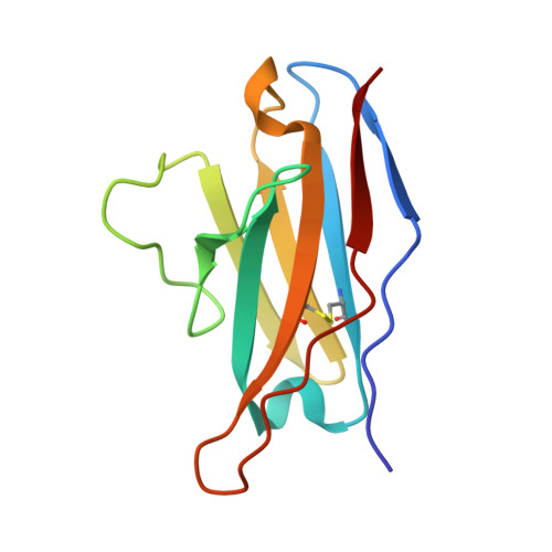
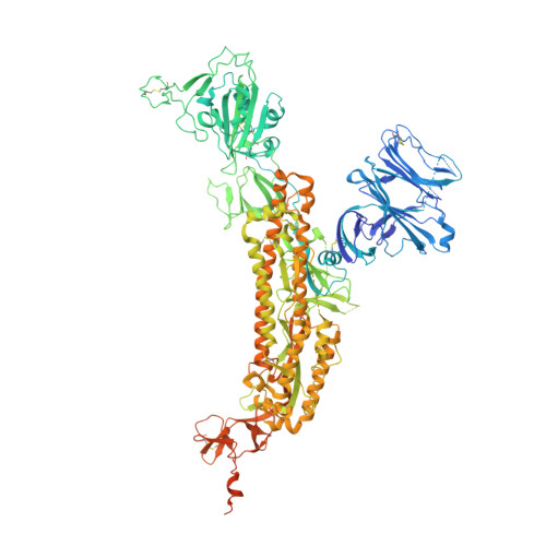
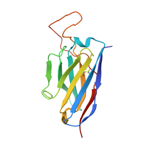
Antibodies targeting a quaternary site on SARS-CoV-2 spike glycoprotein prevent viral receptor engagement by conformational locking.

Liu, L.Casner, R.G.Guo, Y.Wang, Q.Iketani, S.Chan, J.F.Yu, J.Dadonaite, B.Nair, M.S.Mohri, H.Reddem, E.R.Yuan, S.Poon, V.K.Chan, C.C.Yuen, K.Y.Sheng, Z.Huang, Y.Bloom, J.D.Shapiro, L.Ho, D.D.

(2023) Immunity 56: 2442

  • DOI: https://doi.org/10.1016/j.immuni.2023.09.003
  • Primary Citation Related Structures: 
    7UKL, 7UKM

  • PubMed Abstract: 

    SARS-CoV-2 continues to evolve, with many variants evading clinically authorized antibodies. To isolate monoclonal antibodies (mAbs) with broadly neutralizing capacities against the virus, we screened serum samples from convalescing COVID-19 patients. We isolated two mAbs, 12-16 and 12-19, which neutralized all SARS-CoV-2 variants tested, including the XBB subvariants, and prevented infection in hamsters challenged with Omicron BA.1 intranasally. Structurally, both antibodies targeted a conserved quaternary epitope located at the interface between the N-terminal domain and subdomain 1, uncovering a site of vulnerability on SARS-CoV-2 spike. These antibodies prevented viral receptor engagement by locking the receptor-binding domain (RBD) of spike in the down conformation, revealing a mechanism of virus neutralization for non-RBD antibodies. Deep mutational scanning showed that SARS-CoV-2 could mutate to escape 12-19, but such mutations are rarely found in circulating viruses. Antibodies 12-16 and 12-19 hold promise as prophylactic agents for immunocompromised persons who do not respond robustly to COVID-19 vaccines.


  • Organizational Affiliation
    • Aaron Diamond AIDS Research Center, Columbia University Vagelos College of Physicians and Surgeons, New York, NY 10032, USA; Division of Infectious Diseases, Department of Medicine, Columbia University Vagelos College of Physicians and Surgeons, New York, NY 10032, USA. Electronic address: ll3411@cumc.columbia.edu.

Macromolecule Content 

  • Total Structure Weight: 513.06 kDa 
  • Atom Count: 30,234 
  • Modeled Residue Count: 3,795 
  • Deposited Residue Count: 4,539 
  • Unique protein chains: 3

Macromolecules

Find similar proteins by:|  3D Structure
Entity ID: 1
MoleculeChains  Sequence LengthOrganismDetailsImage
12-16 Fab Light ChainA [auth Y],
D [auth L],
I [auth N]
108Homo sapiensMutation(s): 0 
Find similar proteins by:|  3D Structure
Entity ID: 2
MoleculeChains  Sequence LengthOrganismDetailsImage
Spike glycoproteinB,
E [auth C],
F [auth A]
1,273Severe acute respiratory syndrome coronavirus 2Mutation(s): 1 
Gene Names: S2
UniProt
Find proteins for P0DTC2 (Severe acute respiratory syndrome coronavirus 2)
Explore P0DTC2 
Go to UniProtKB:  P0DTC2
Entity Groups
Sequence Clusters30% Identity50% Identity70% Identity90% Identity95% Identity100% Identity
UniProt GroupP0DTC2
Glycosylation
Glycosylation Sites: 16Go to GlyGen: P0DTC2-1
Sequence Annotations
Expand
Reference Sequence
Find similar proteins by:|  3D Structure
Entity ID: 3
MoleculeChains  Sequence LengthOrganismDetailsImage
12-16 Fab Heavy ChainC [auth X],
G [auth H],
H [auth M]
132Homo sapiensMutation(s): 0 

Small Molecules

Ligands 1 Unique
IDChains Name / Formula / InChI Key2D Diagram3D Interactions
NAG

Query on NAG


Download:Ideal Coordinates CCD File
AA [auth C]
AB [auth A]
BA [auth C]
BB [auth A]
CA [auth C]
AA [auth C],
AB [auth A],
BA [auth C],
BB [auth A],
CA [auth C],
CB [auth A],
DA [auth C],
DB [auth A],
EA [auth C],
EB [auth A],
FA [auth C],
GA [auth C],
HA [auth C],
IA [auth C],
J [auth B],
JA [auth C],
K [auth B],
KA [auth C],
L [auth B],
LA [auth C],
M [auth B],
MA [auth C],
N [auth B],
NA [auth C],
O [auth B],
OA [auth C],
P [auth B],
PA [auth A],
Q [auth B],
QA [auth A],
R [auth B],
RA [auth A],
S [auth B],
SA [auth A],
T [auth B],
TA [auth A],
U [auth B],
UA [auth A],
V [auth B],
VA [auth A],
W [auth B],
WA [auth A],
X [auth B],
XA [auth A],
Y [auth B],
YA [auth A],
Z [auth C],
ZA [auth A]
2-acetamido-2-deoxy-beta-D-glucopyranose
C8 H15 N O6
OVRNDRQMDRJTHS-FMDGEEDCSA-N

Experimental Data & Validation

Experimental Data

  • Method: ELECTRON MICROSCOPY
  • Resolution: 3.09 Å
  • Aggregation State: PARTICLE 
  • Reconstruction Method: SINGLE PARTICLE 
EM Software:
TaskSoftware PackageVersion
RECONSTRUCTIONcryoSPARC3.3

Structure Validation

View Full Validation Report


Entry History 

& Funding Information

Deposition Data


Funding OrganizationLocationGrant Number
Jack Ma FoundationChina--

Revision History  (Full details and data files)

  • Version 1.0: 2023-10-04
    Type: Initial release
  • Version 1.1: 2023-10-11
    Changes: Database references
  • Version 1.2: 2023-10-25
    Changes: Database references
  • Version 1.3: 2024-10-30
    Changes: Data collection, Structure summary