7U6V

Cryo-EM structure of Shiga toxin 2 in complex with the native ribosomal P-stalk


Experimental Data Snapshot

  • Method: ELECTRON MICROSCOPY
  • Resolution: 4.10 Å
  • Aggregation State: PARTICLE 
  • Reconstruction Method: SINGLE PARTICLE 

wwPDB Validation   3D Report Full Report


This is version 1.0 of the entry. See complete history


Literature

Cryo-EM structure of Shiga toxin 2 in complex with the native ribosomal P-stalk reveals residues involved in the binding interaction.

Kulczyk, A.W.Sorzano, C.O.S.Grela, P.Tchorzewski, M.Tumer, N.E.Li, X.P.

(2022) J Biol Chem 299: 102795-102795

  • DOI: https://doi.org/10.1016/j.jbc.2022.102795
  • Primary Citation of Related Structures:  
    7U6V

  • PubMed Abstract: 

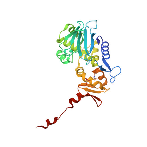
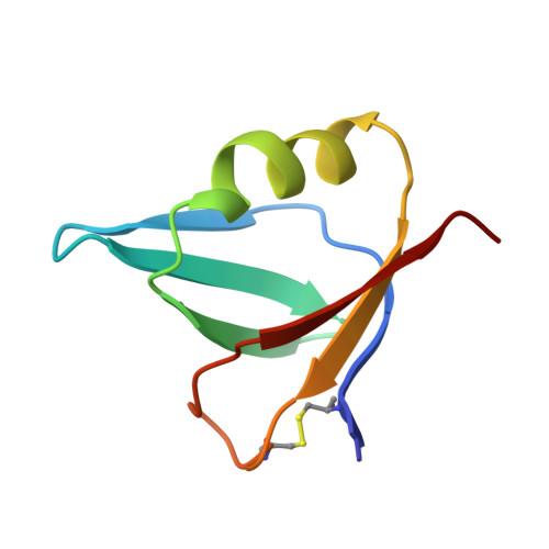
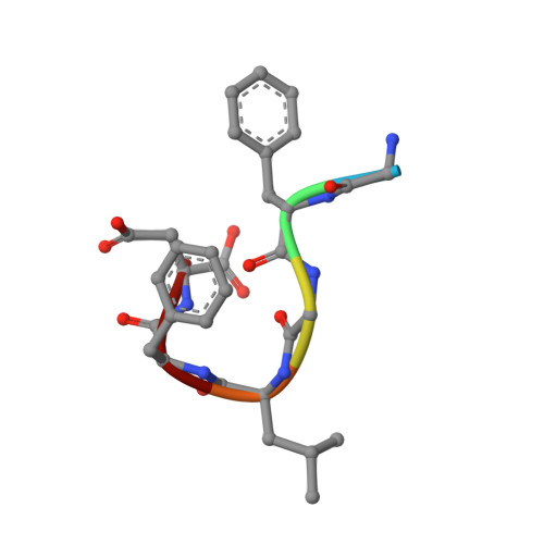
    Shiga toxin 2a (Stx2a) is the virulence factor of enterohemorrhagic Escherichia coli. The catalytic A1 subunit of Stx2a (Stx2A1) interacts with the ribosomal P-stalk for loading onto the ribosome and depurination of the sarcin-ricin loop, which halts protein synthesis. Because of the intrinsic flexibility of the P-stalk, a structure of the Stx2a-P-stalk complex is currently unknown. We demonstrated that the native P-stalk pentamer binds to Stx2a with nanomolar affinity, and we employed cryo-EM to determine a structure of the 72 kDa Stx2a complexed with the P-stalk. The structure identifies Stx2A1 residues involved in binding and reveals that Stx2a is anchored to the P-stalk via only the last six amino acids from the C-terminal domain of a single P-protein. For the first time, the cryo-EM structure shows the loop connecting Stx2A1 and Stx2A2, which is critical for activation of the toxin. Our principal component analysis of the cryo-EM data reveals the intrinsic dynamics of the Stx2a-P-stalk interaction, including conformational changes in the P-stalk binding site occurring upon complex formation. Our computational analysis unveils the propensity for structural rearrangements within the C-terminal domain, with its C-terminal six amino acids transitioning from a random coil to an α-helix upon binding to Stx2a. In conclusion, our cryo-EM structure sheds new light into the dynamics of the Stx2a-P-stalk interaction and indicates that the binding interface between Stx2a and the P-stalk is the potential target for drug discovery.


  • Organizational Affiliation

    Institute for Quantitative Biomedicine, Department of Biochemistry and Microbiology, Rutgers University, Piscataway, New Jersey, USA. Electronic address: arek.kulczyk@rutgers.edu.


Macromolecules
Find similar proteins by:  (by identity cutoff)  |  3D Structure
Entity ID: 1
MoleculeChains Sequence LengthOrganismDetailsImage
Shiga toxin 2a subunit A (Stx2A)297Shigella dysenteriaeMutation(s): 0 
Gene Names: stxA2
EC: 3.2.2.22
UniProt
Find proteins for Q7DI68 (Escherichia coli O157:H7)
Explore Q7DI68 
Go to UniProtKB:  Q7DI68
Entity Groups  
Sequence Clusters30% Identity50% Identity70% Identity90% Identity95% Identity100% Identity
UniProt GroupQ7DI68
Sequence Annotations
Expand
  • Reference Sequence
Find similar proteins by:  (by identity cutoff)  |  3D Structure
Entity ID: 2
MoleculeChains Sequence LengthOrganismDetailsImage
Shiga toxin 2a subunit B (Stx2B)
B, C, D, E, F
70Shigella dysenteriaeMutation(s): 0 
UniProt
Find proteins for A7UQX3 (Escherichia coli O157:H7)
Explore A7UQX3 
Go to UniProtKB:  A7UQX3
Entity Groups  
Sequence Clusters30% Identity50% Identity70% Identity90% Identity95% Identity100% Identity
UniProt GroupA7UQX3
Sequence Annotations
Expand
  • Reference Sequence

Find similar proteins by:  Sequence   |   3D Structure  

Entity ID: 3
MoleculeChains Sequence LengthOrganismDetailsImage
C-terminal domain (CTD) from the Ribosomal P-stalkG [auth P]6Saccharomyces cerevisiaeMutation(s): 0 
UniProt
Find proteins for P05317 (Saccharomyces cerevisiae (strain ATCC 204508 / S288c))
Explore P05317 
Go to UniProtKB:  P05317
Entity Groups  
UniProt GroupP05317
Sequence Annotations
Expand
  • Reference Sequence
Experimental Data & Validation

Experimental Data

  • Method: ELECTRON MICROSCOPY
  • Resolution: 4.10 Å
  • Aggregation State: PARTICLE 
  • Reconstruction Method: SINGLE PARTICLE 
EM Software:
TaskSoftware PackageVersion
MODEL REFINEMENTPHENIX1.18

Structure Validation

View Full Validation Report


Entry History & Funding Information

Deposition Data


Funding OrganizationLocationGrant Number
National Institutes of Health/National Institute Of Allergy and Infectious Diseases (NIH/NIAID)United StatesAI141635

Revision History  (Full details and data files)

  • Version 1.0: 2023-01-11
    Type: Initial release