7TVD

Crystal structure of the kinase domain of EGFR exon-19 (del-747-749) mutant


Experimental Data Snapshot

  • Method: X-RAY DIFFRACTION
  • Resolution: 2.96 Å
  • R-Value Free: 0.277 
  • R-Value Work: 0.240 
  • R-Value Observed: 0.242 

wwPDB Validation   3D Report Full Report


This is version 1.3 of the entry. See complete history


Literature

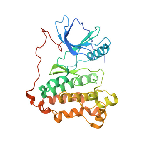
Biochemical and structural basis for differential inhibitor sensitivity of EGFR with distinct exon 19 mutations.

van Alderwerelt van Rosenburgh, I.K.Lu, D.M.Grant, M.J.Stayrook, S.E.Phadke, M.Walther, Z.Goldberg, S.B.Politi, K.Lemmon, M.A.Ashtekar, K.D.Tsutsui, Y.

(2022) Nat Commun 13: 6791-6791

  • DOI: https://doi.org/10.1038/s41467-022-34398-z
  • Primary Citation of Related Structures:  
    7TVD

  • PubMed Abstract: 

    Tyrosine kinase inhibitors (TKIs) are used to treat non-small cell lung cancers (NSCLC) driven by epidermal growth factor receptor (EGFR) mutations in the tyrosine kinase domain (TKD). TKI responses vary across tumors driven by the heterogeneous group of exon 19 deletions and mutations, but the molecular basis for these differences is not understood. Using purified TKDs, we compared kinetic properties of several exon 19 variants. Although unaltered for the second generation TKI afatinib, sensitivity varied significantly for both the first and third generation TKIs erlotinib and osimertinib. The most sensitive variants showed reduced ATP-binding affinity, whereas those associated with primary resistance retained wild type ATP-binding characteristics (and low K M, ATP ). Through crystallographic and hydrogen-deuterium exchange mass spectrometry (HDX-MS) studies, we identify possible origins for the altered ATP-binding affinity underlying TKI sensitivity and resistance, and propose a basis for classifying uncommon exon 19 variants that may have predictive clinical value.


  • Organizational Affiliation

    Department of Pharmacology, Yale University School of Medicine, New Haven, CT, 06520, USA.


Macromolecules
Find similar proteins by:  (by identity cutoff)  |  3D Structure
Entity ID: 1
MoleculeChains Sequence LengthOrganismDetailsImage
Epidermal growth factor receptor335Homo sapiensMutation(s): 0 
Gene Names: EGFRERBBERBB1HER1
EC: 2.7.10.1
UniProt & NIH Common Fund Data Resources
Find proteins for P00533 (Homo sapiens)
Explore P00533 
Go to UniProtKB:  P00533
PHAROS:  P00533
GTEx:  ENSG00000146648 
Entity Groups  
Sequence Clusters30% Identity50% Identity70% Identity90% Identity95% Identity100% Identity
UniProt GroupP00533
Sequence Annotations
Expand
  • Reference Sequence
Experimental Data & Validation

Experimental Data

  • Method: X-RAY DIFFRACTION
  • Resolution: 2.96 Å
  • R-Value Free: 0.277 
  • R-Value Work: 0.240 
  • R-Value Observed: 0.242 
  • Space Group: I 2 3
Unit Cell:
Length ( Å )Angle ( ˚ )
a = 149.42α = 90
b = 149.42β = 90
c = 149.42γ = 90
Software Package:
Software NamePurpose
PHENIXrefinement
XDSdata reduction
PDB_EXTRACTdata extraction
SCALAdata scaling
PHASERphasing

Structure Validation

View Full Validation Report


Entry History & Funding Information

Deposition Data


Funding OrganizationLocationGrant Number
National Institutes of Health/National Cancer Institute (NIH/NCI)United StatesGR110199

Revision History  (Full details and data files)

  • Version 1.0: 2022-02-16
    Type: Initial release
  • Version 1.1: 2022-08-31
    Changes: Database references
  • Version 1.2: 2022-11-30
    Changes: Database references
  • Version 1.3: 2023-10-25
    Changes: Data collection, Refinement description