7TTH | pdb_00007tth

Human potassium-chloride cotransporter 1 in inward-open state


Experimental Data Snapshot

  • Method: ELECTRON MICROSCOPY
  • Resolution: 3.25 Å
  • Aggregation State: PARTICLE 
  • Reconstruction Method: SINGLE PARTICLE 

wwPDB Validation 3D Report Full Report

Validation slider image for 7TTH

This is version 1.3 of the entry. See complete history

Literature

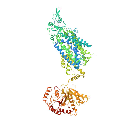
Structure of the human cation-chloride cotransport KCC1 in an outward-open state.

Zhao, Y.Shen, J.Wang, Q.Ruiz Munevar, M.J.Vidossich, P.De Vivo, M.Zhou, M.Cao, E.

(2022) Proc Natl Acad Sci U S A 119: e2109083119-e2109083119

  • DOI: https://doi.org/10.1073/pnas.2109083119
  • Primary Citation Related Structures: 
    7TTH, 7TTI

  • PubMed Abstract: 

    Cation-chloride cotransporters (CCCs) catalyze electroneutral symport of Cl - with Na + and/or K + across membranes. CCCs are fundamental in cell volume homeostasis, transepithelia ion movement, maintenance of intracellular Cl - concentration, and neuronal excitability. Here, we present a cryoelectron microscopy structure of human K + -Cl - cotransporter (KCC)1 bound with the VU0463271 inhibitor in an outward-open state. In contrast to many other amino acid-polyamine-organocation transporter cousins, our first outward-open CCC structure reveals that opening the KCC1 extracellular ion permeation path does not involve hinge-bending motions of the transmembrane (TM) 1 and TM6 half-helices. Instead, rocking of TM3 and TM8, together with displacements of TM4, TM9, and a conserved intracellular loop 1 helix, underlie alternate opening and closing of extracellular and cytoplasmic vestibules. We show that KCC1 intriguingly exists in one of two distinct dimeric states via different intersubunit interfaces. Our studies provide a blueprint for understanding the mechanisms of CCCs and their inhibition by small molecule compounds.


  • Organizational Affiliation
    • Department of Biochemistry, University of Utah School of Medicine, Salt Lake City, UT 84112.

Macromolecule Content 

  • Total Structure Weight: 243.32 kDa 
  • Atom Count: 12,698 
  • Modeled Residue Count: 1,750 
  • Deposited Residue Count: 2,170 
  • Unique protein chains: 1

Macromolecules

Find similar proteins by:|  3D Structure
Entity ID: 1
MoleculeChains  Sequence LengthOrganismDetailsImage
Solute carrier family 12 member 4
A, B
1,085Homo sapiensMutation(s): 0 
Gene Names: SLC12A4KCC1
Membrane Entity: Yes 
UniProt & NIH Common Fund Data Resources
Find proteins for Q9UP95 (Homo sapiens)
Explore Q9UP95 
Go to UniProtKB:  Q9UP95
PHAROS:  Q9UP95
GTEx:  ENSG00000124067 
Entity Groups
Sequence Clusters30% Identity50% Identity70% Identity90% Identity95% Identity100% Identity
UniProt GroupQ9UP95
Glycosylation
Glycosylation Sites: 2Go to GlyGen: Q9UP95-1
Sequence Annotations
Expand
Reference Sequence

Oligosaccharides

Help  
Entity ID: 2
MoleculeChains Length2D Diagram GlycosylationD Interactions
2-acetamido-2-deoxy-beta-D-glucopyranose-(1-4)-2-acetamido-2-deoxy-beta-D-glucopyranose
C, D, E, F
2N-Glycosylation
Glycosylation Resources
GlyTouCan: G42666HT
GlyCosmos: G42666HT
GlyGen: G42666HT

Experimental Data & Validation

Experimental Data

  • Method: ELECTRON MICROSCOPY
  • Resolution: 3.25 Å
  • Aggregation State: PARTICLE 
  • Reconstruction Method: SINGLE PARTICLE 
EM Software:
TaskSoftware PackageVersion
MODEL REFINEMENTPHENIX

Structure Validation

View Full Validation Report


Entry History 

& Funding Information

Deposition Data


Funding OrganizationLocationGrant Number
Not funded--

Revision History  (Full details and data files)

  • Version 1.0: 2022-06-29
    Type: Initial release
  • Version 1.1: 2022-07-13
    Changes: Database references
  • Version 1.2: 2024-10-09
    Changes: Data collection, Structure summary
  • Version 1.3: 2025-05-28
    Changes: Data collection