7TLM

Structure of Atopobium parvulum SufS


Experimental Data Snapshot

  • Method: X-RAY DIFFRACTION
  • Resolution: 1.80 Å
  • R-Value Free: 0.225 
  • R-Value Work: 0.195 
  • R-Value Observed: 0.196 

wwPDB Validation   3D Report Full Report


Ligand Structure Quality Assessment 


This is version 2.2 of the entry. See complete history


Literature

Structural analysis of Atopobium parvulum SufS cysteine desulfurase linked to Crohn's disease.

Karunakaran, G.Yang, Y.Tremblay, V.Ning, Z.Martin, J.Belaouad, A.Figeys, D.Brunzelle, J.S.Giguere, P.M.Stintzi, A.Couture, J.F.

(2022) FEBS Lett 596: 898-909

  • DOI: https://doi.org/10.1002/1873-3468.14295
  • Primary Citation of Related Structures:  
    7TLM, 7TLP, 7TLQ, 7TLR

  • PubMed Abstract: 

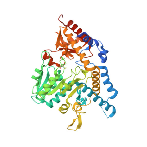
    Crohn's disease (CD) is characterized by the chronic inflammation of the gastrointestinal tract. A dysbiotic microbiome and a defective immune system are linked to CD, where hydrogen sulfide (H 2 S) microbial producers positively correlate with the severity of the disease. Atopobium parvulum is a key H 2 S producer from the microbiome of CD patients. In this study, the biochemical characterization of two Atopobium parvulum cysteine desulfurases, ApSufS and ApCsdB, shows that the enzymes are allosterically regulated. Structural analyses reveal that ApSufS forms a dimer with conserved characteristics observed in type II cysteine desulfurases. Four residues surrounding the active site are essential to catalyse cysteine desulfurylation, and a segment of short-chain residues grant access for substrate binding. A better understanding of ApSufS will help future avenues for CD treatment.


  • Organizational Affiliation

    Department of Biochemistry, Microbiology and Immunology, Faculty of Medicine, University of Ottawa, Canada.


Macromolecules
Find similar proteins by:  (by identity cutoff)  |  3D Structure
Entity ID: 1
MoleculeChains Sequence LengthOrganismDetailsImage
Cysteine desulfurase447Lancefieldella parvulaMutation(s): 0 
Gene Names: Apar_0399
EC: 2.8.1.7
UniProt
Find proteins for C8W9P2 (Lancefieldella parvula (strain ATCC 33793 / DSM 20469 / CCUG 32760 / JCM 10300 / KCTC 3663 / VPI 0546 / 1246))
Explore C8W9P2 
Go to UniProtKB:  C8W9P2
Entity Groups  
Sequence Clusters30% Identity50% Identity70% Identity90% Identity95% Identity100% Identity
UniProt GroupC8W9P2
Sequence Annotations
Expand
  • Reference Sequence
Small Molecules
Ligands 1 Unique
IDChains Name / Formula / InChI Key2D Diagram3D Interactions
PLP (Subject of Investigation/LOI)
Query on PLP

Download Ideal Coordinates CCD File 
B [auth A]PYRIDOXAL-5'-PHOSPHATE
C8 H10 N O6 P
NGVDGCNFYWLIFO-UHFFFAOYSA-N
Modified Residues  1 Unique
IDChains TypeFormula2D DiagramParent
CSS
Query on CSS
A
L-PEPTIDE LINKINGC3 H7 N O2 S2CYS
Experimental Data & Validation

Experimental Data

  • Method: X-RAY DIFFRACTION
  • Resolution: 1.80 Å
  • R-Value Free: 0.225 
  • R-Value Work: 0.195 
  • R-Value Observed: 0.196 
  • Space Group: P 61 2 2
Unit Cell:
Length ( Å )Angle ( ˚ )
a = 92.011α = 90
b = 92.011β = 90
c = 178.242γ = 120
Software Package:
Software NamePurpose
PHENIXrefinement
d*TREKdata scaling
PDB_EXTRACTdata extraction
d*TREKdata reduction
PHASERphasing
Cootmodel building

Structure Validation

View Full Validation Report


Ligand Structure Quality Assessment 


Entry History & Funding Information

Deposition Data


Funding OrganizationLocationGrant Number
Canadian Institutes of Health Research (CIHR)Canada--

Revision History  (Full details and data files)

  • Version 1.0: 2022-02-16
    Type: Initial release
  • Version 1.1: 2022-04-27
    Changes: Database references
  • Version 2.0: 2022-07-06
    Type: Coordinate replacement
    Reason: Ligand identity
    Changes: Advisory, Atomic model, Author supporting evidence, Data collection, Database references, Derived calculations, Polymer sequence, Refinement description, Source and taxonomy, Structure summary
  • Version 2.1: 2023-10-18
    Changes: Data collection, Refinement description
  • Version 2.2: 2023-11-15
    Changes: Data collection