7TJ9 | pdb_00007tj9

Cryo-EM structure of the human Nax channel in complex with beta3 solved in GDN


Experimental Data Snapshot

  • Method: ELECTRON MICROSCOPY
  • Resolution: 2.90 Å
  • Aggregation State: PARTICLE 
  • Reconstruction Method: SINGLE PARTICLE 

wwPDB Validation 3D Report Full Report

Validation slider image for 7TJ9

This is version 1.2 of the entry. See complete history

Literature

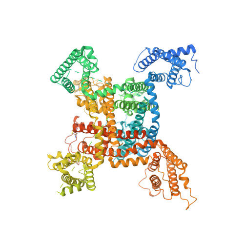
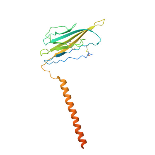
Structure-guided unlocking of Na X reveals a non-selective tetrodotoxin-sensitive cation channel.

Noland, C.L.Chua, H.C.Kschonsak, M.Heusser, S.A.Braun, N.Chang, T.Tam, C.Tang, J.Arthur, C.P.Ciferri, C.Pless, S.A.Payandeh, J.

(2022) Nat Commun 13: 1416-1416

  • DOI: https://doi.org/10.1038/s41467-022-28984-4
  • Primary Citation Related Structures: 
    7TJ8, 7TJ9

  • PubMed Abstract: 

    Unlike classical voltage-gated sodium (Na V ) channels, Na X has been characterized as a voltage-insensitive, tetrodotoxin-resistant, sodium (Na + )-activated channel involved in regulating Na + homeostasis. However, Na X remains refractory to functional characterization in traditional heterologous systems. Here, to gain insight into its atypical physiology, we determine structures of the human Na X channel in complex with the auxiliary β3-subunit. Na X reveals structural alterations within the selectivity filter, voltage sensor-like domains, and pore module. We do not identify an extracellular Na + -sensor or any evidence for a Na + -based activation mechanism in Na X . Instead, the S6-gate remains closed, membrane lipids fill the central cavity, and the domain III-IV linker restricts S6-dilation. We use protein engineering to identify three pore-wetting mutations targeting the hydrophobic S6-gate that unlock a robust voltage-insensitive leak conductance. This constitutively active Na X -QTT channel construct is non-selective among monovalent cations, inhibited by extracellular calcium, and sensitive to classical Na V channel blockers, including tetrodotoxin. Our findings highlight a functional diversity across the Na V channel scaffold, reshape our understanding of Na X physiology, and provide a template to demystify recalcitrant ion channels.


  • Organizational Affiliation
    • Department of Structural Biology, Genentech Inc., South San Francisco, CA, 94080, USA.

Macromolecule Content 

  • Total Structure Weight: 234.37 kDa 
  • Atom Count: 10,975 
  • Modeled Residue Count: 1,273 
  • Deposited Residue Count: 1,952 
  • Unique protein chains: 2

Macromolecules

Find similar proteins by:|  3D Structure
Entity ID: 1
MoleculeChains  Sequence LengthOrganismDetailsImage
Sodium channel protein type 7 subunit alpha1,737Homo sapiensMutation(s): 0 
Gene Names: SCN7ASCN6A
Membrane Entity: Yes 
UniProt & NIH Common Fund Data Resources
Find proteins for Q01118 (Homo sapiens)
Explore Q01118 
Go to UniProtKB:  Q01118
PHAROS:  Q01118
GTEx:  ENSG00000136546 
Entity Groups
Sequence Clusters30% Identity50% Identity70% Identity90% Identity95% Identity100% Identity
UniProt GroupQ01118
Glycosylation
Glycosylation Sites: 2Go to GlyGen: Q01118-1
Sequence Annotations
Expand
Reference Sequence
Find similar proteins by:|  3D Structure
Entity ID: 2
MoleculeChains  Sequence LengthOrganismDetailsImage
Sodium channel subunit beta-3215Homo sapiensMutation(s): 0 
Gene Names: SCN3BKIAA1158
Membrane Entity: Yes 
UniProt & NIH Common Fund Data Resources
Find proteins for Q9NY72 (Homo sapiens)
Explore Q9NY72 
Go to UniProtKB:  Q9NY72
PHAROS:  Q9NY72
GTEx:  ENSG00000166257 
Entity Groups
Sequence Clusters30% Identity50% Identity70% Identity90% Identity95% Identity100% Identity
UniProt GroupQ9NY72
Glycosylation
Glycosylation Sites: 4Go to GlyGen: Q9NY72-1
Sequence Annotations
Expand
Reference Sequence

Oligosaccharides

Help  
Entity ID: 3
MoleculeChains Length2D Diagram GlycosylationD Interactions
2-acetamido-2-deoxy-beta-D-glucopyranose-(1-4)-2-acetamido-2-deoxy-beta-D-glucopyranose
C
2N-Glycosylation
Glycosylation Resources
GlyTouCan: G42666HT
GlyCosmos: G42666HT
GlyGen: G42666HT

Small Molecules

Ligands 6 Unique
IDChains Name / Formula / InChI Key2D Diagram3D Interactions
Q7G

Query on Q7G


Download:Ideal Coordinates CCD File
U [auth A]2-{[(4-O-alpha-D-glucopyranosyl-alpha-D-glucopyranosyl)oxy]methyl}-4-{[(3beta,9beta,14beta,17beta,25R)-spirost-5-en-3-yl]oxy}butyl 4-O-alpha-D-glucopyranosyl-alpha-D-glucopyranoside
C56 H92 O25
LKBFXDNKNZXHHW-NXLTVWPKSA-N
POV

Query on POV


Download:Ideal Coordinates CCD File
E [auth A],
F [auth A],
K [auth A]
(2S)-3-(hexadecanoyloxy)-2-[(9Z)-octadec-9-enoyloxy]propyl 2-(trimethylammonio)ethyl phosphate
C42 H82 N O8 P
WTJKGGKOPKCXLL-PFDVCBLKSA-N
PEV

Query on PEV


Download:Ideal Coordinates CCD File
D [auth A],
H [auth A],
N [auth A]
(1S)-2-{[(2-AMINOETHOXY)(HYDROXY)PHOSPHORYL]OXY}-1-[(PALMITOYLOXY)METHYL]ETHYL STEARATE
C39 H78 N O8 P
RPJZYOHZALDGKI-QNGWXLTQSA-N
CLR

Query on CLR


Download:Ideal Coordinates CCD File
S [auth A],
T [auth A]
CHOLESTEROL
C27 H46 O
HVYWMOMLDIMFJA-DPAQBDIFSA-N
PLM

Query on PLM


Download:Ideal Coordinates CCD File
G [auth A]
I [auth A]
J [auth A]
L [auth A]
M [auth A]
G [auth A],
I [auth A],
J [auth A],
L [auth A],
M [auth A],
P [auth A],
Q [auth A],
R [auth A]
PALMITIC ACID
C16 H32 O2
IPCSVZSSVZVIGE-UHFFFAOYSA-N
NAG

Query on NAG


Download:Ideal Coordinates CCD File
O [auth A],
V [auth B],
W [auth B],
X [auth B],
Y [auth B]
2-acetamido-2-deoxy-beta-D-glucopyranose
C8 H15 N O6
OVRNDRQMDRJTHS-FMDGEEDCSA-N

Experimental Data & Validation

Experimental Data

  • Method: ELECTRON MICROSCOPY
  • Resolution: 2.90 Å
  • Aggregation State: PARTICLE 
  • Reconstruction Method: SINGLE PARTICLE 
EM Software:
TaskSoftware PackageVersion
MODEL REFINEMENTPHENIX

Structure Validation

View Full Validation Report


Entry History 

& Funding Information

Deposition Data


Funding OrganizationLocationGrant Number
Not funded--

Revision History  (Full details and data files)

  • Version 1.0: 2022-03-30
    Type: Initial release
  • Version 1.1: 2024-10-09
    Changes: Data collection, Structure summary
  • Version 1.2: 2025-05-28
    Changes: Data collection