7TJ8 | pdb_00007tj8

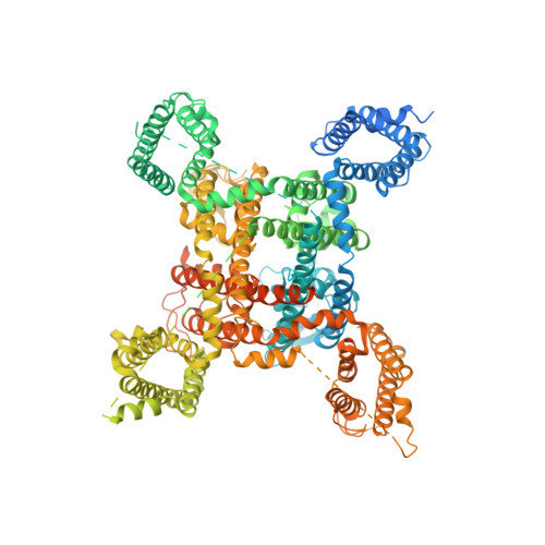
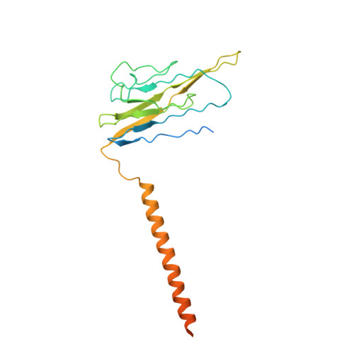
Cryo-EM structure of the human Nax channel in complex with beta3 solved in nanodiscs


Experimental Data Snapshot

  • Method: ELECTRON MICROSCOPY
  • Resolution: 3.20 Å
  • Aggregation State: PARTICLE 
  • Reconstruction Method: SINGLE PARTICLE 

wwPDB Validation 3D Report Full Report

Validation slider image for 7TJ8

This is version 1.2 of the entry. See complete history. 

Literature

Structure-guided unlocking of Na X reveals a non-selective tetrodotoxin-sensitive cation channel.

Noland, C.L., Chua, H.C., Kschonsak, M., Heusser, S.A., Braun, N., Chang, T., Tam, C., Tang, J., Arthur, C.P., Ciferri, C., Pless, S.A., Payandeh, J.

(2022) Nat Commun 13: 1416-1416

  • DOI: https://doi.org/10.1038/s41467-022-28984-4
  • Primary Citation Related Structures: 
    7TJ8, 7TJ9

  • PubMed Abstract: 

    Unlike classical voltage-gated sodium (Na V ) channels, Na X has been characterized as a voltage-insensitive, tetrodotoxin-resistant, sodium (Na + )-activated channel involved in regulating Na + homeostasis. However, Na X remains refractory to functional characterization in traditional heterologous systems. Here, to gain insight into its atypical physiology, we determine structures of the human Na X channel in complex with the auxiliary β3-subunit. Na X reveals structural alterations within the selectivity filter, voltage sensor-like domains, and pore module. We do not identify an extracellular Na + -sensor or any evidence for a Na + -based activation mechanism in Na X . Instead, the S6-gate remains closed, membrane lipids fill the central cavity, and the domain III-IV linker restricts S6-dilation. We use protein engineering to identify three pore-wetting mutations targeting the hydrophobic S6-gate that unlock a robust voltage-insensitive leak conductance. This constitutively active Na X -QTT channel construct is non-selective among monovalent cations, inhibited by extracellular calcium, and sensitive to classical Na V channel blockers, including tetrodotoxin. Our findings highlight a functional diversity across the Na V channel scaffold, reshape our understanding of Na X physiology, and provide a template to demystify recalcitrant ion channels.


  • Organizational Affiliation: 
    • Department of Structural Biology, Genentech Inc., South San Francisco, CA, 94080, USA.

Macromolecule Content 

  • Total Structure Weight: 231.88 kDa 
  • Atom Count: 10,551 
  • Modeled Residue Count: 1,242 
  • Deposited Residue Count: 1,952 
  • Unique protein chains: 2

Macromolecules

Find similar proteins by:|  3D Structure
Entity ID: 1
MoleculeChains  Sequence LengthOrganismDetailsImage
Sodium channel protein type 7 subunit alpha1,737Homo sapiensMutation(s): 0 
Gene Names: SCN7A, SCN6A
Membrane Entity: Yes 
UniProt & NIH Common Fund Data Resources
Find proteins for Q01118 (Homo sapiens)
Explore Q01118 
Go to UniProtKB:  Q01118
PHAROS:  Q01118
GTEx:  ENSG00000136546 
Entity Groups
Sequence Clusters30% Identity50% Identity70% Identity90% Identity95% Identity100% Identity
UniProt GroupQ01118
Glycosylation
Glycosylation Sites: 1Go to GlyGen: Q01118-1
Sequence Annotations
Expand
Reference Sequence
Find similar proteins by:|  3D Structure
Entity ID: 2
MoleculeChains  Sequence LengthOrganismDetailsImage
Sodium channel subunit beta-3215Homo sapiensMutation(s): 0 
Gene Names: SCN3B, KIAA1158
Membrane Entity: Yes 
UniProt & NIH Common Fund Data Resources
Find proteins for Q9NY72 (Homo sapiens)
Explore Q9NY72 
Go to UniProtKB:  Q9NY72
PHAROS:  Q9NY72
GTEx:  ENSG00000166257 
Entity Groups
Sequence Clusters30% Identity50% Identity70% Identity90% Identity95% Identity100% Identity
UniProt GroupQ9NY72
Glycosylation
Glycosylation Sites: 1Go to GlyGen: Q9NY72-1
Sequence Annotations
Expand
Reference Sequence

Small Molecules

Ligands 5 Unique
IDChains Name / Formula / InChI Key2D Diagram3D Interactions
POV

Query on POV


Download:Ideal Coordinates CCD File
D [auth A],
E [auth A],
J [auth A]
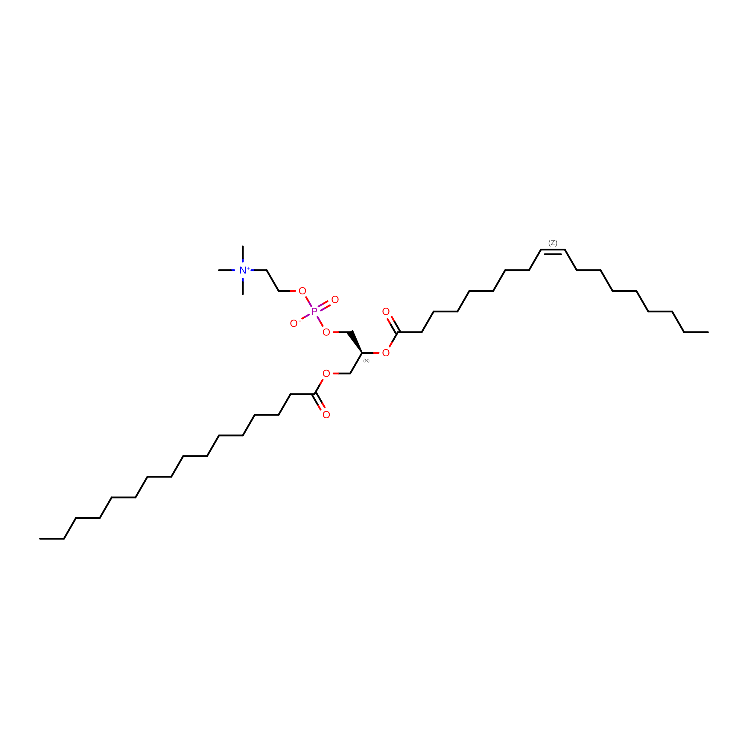
(2S)-3-(hexadecanoyloxy)-2-[(9Z)-octadec-9-enoyloxy]propyl 2-(trimethylammonio)ethyl phosphate
C42 H82 N O8 P
WTJKGGKOPKCXLL-PFDVCBLKSA-N
PEV

Query on PEV


Download:Ideal Coordinates CCD File
C [auth A],
G [auth A],
O [auth A]
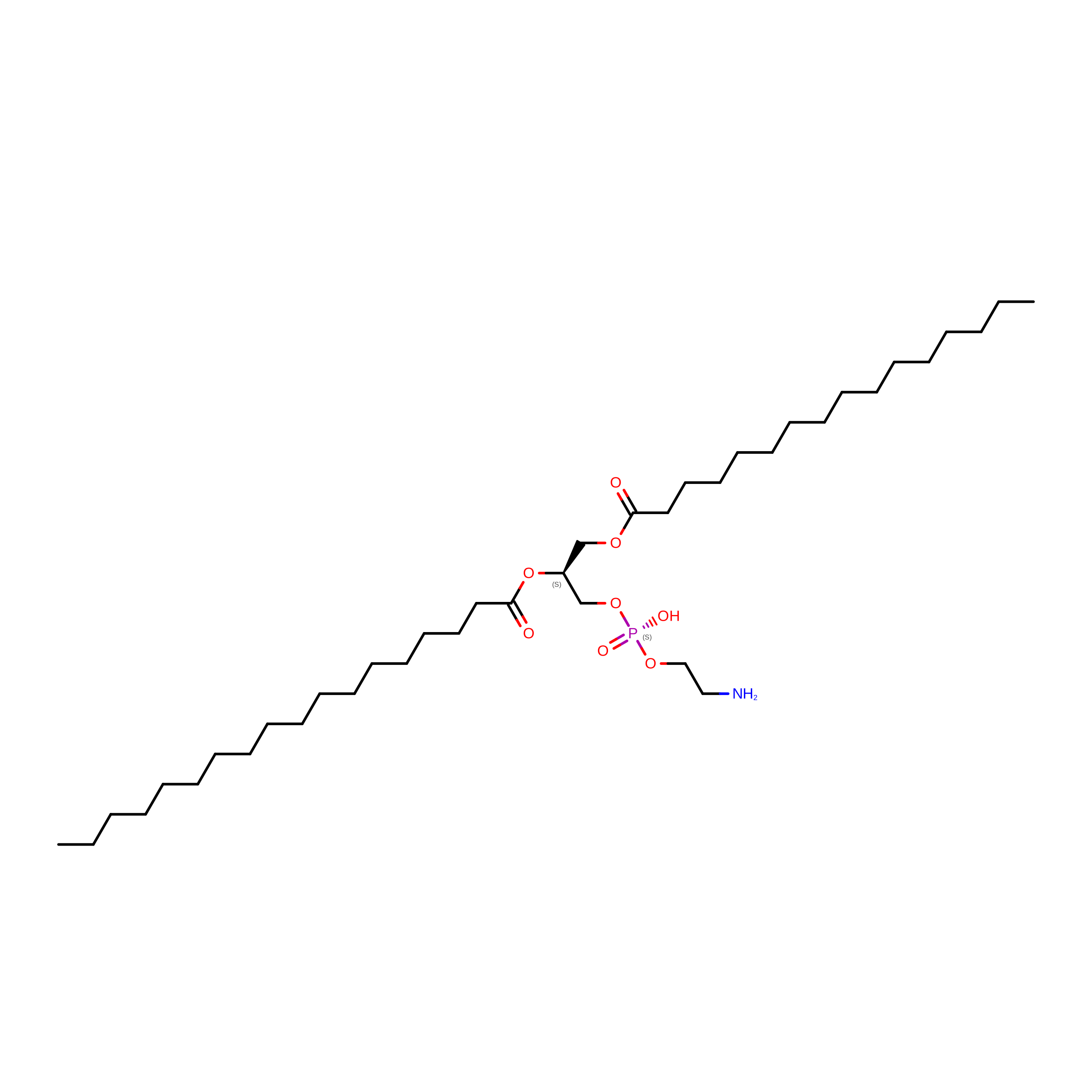
(1S)-2-{[(2-AMINOETHOXY)(HYDROXY)PHOSPHORYL]OXY}-1-[(PALMITOYLOXY)METHYL]ETHYL STEARATE
C39 H78 N O8 P
RPJZYOHZALDGKI-QNGWXLTQSA-N
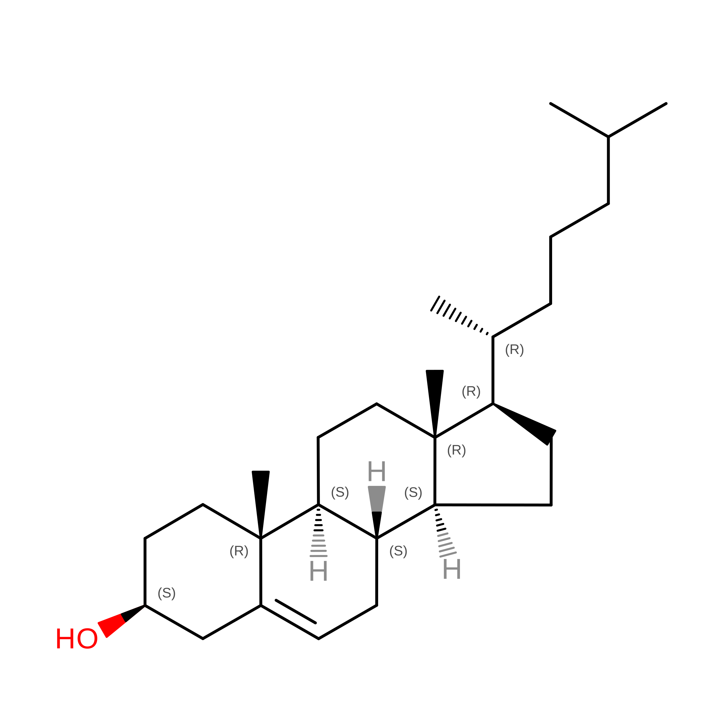
CLR

Query on CLR


Download:Ideal Coordinates CCD File
Q [auth A]CHOLESTEROL
C27 H46 O
HVYWMOMLDIMFJA-DPAQBDIFSA-N
PLM

Query on PLM


Download:Ideal Coordinates CCD File
F [auth A]
H [auth A]
I [auth A]
K [auth A]
L [auth A]
F [auth A],
H [auth A],
I [auth A],
K [auth A],
L [auth A],
M [auth A]
PALMITIC ACID
C16 H32 O2
IPCSVZSSVZVIGE-UHFFFAOYSA-N
NAG

Query on NAG


Download:Ideal Coordinates CCD File
N [auth A],
P [auth A],
R [auth B],
S [auth B],
T [auth B]
2-acetamido-2-deoxy-beta-D-glucopyranose
C8 H15 N O6
OVRNDRQMDRJTHS-FMDGEEDCSA-N

Experimental Data & Validation

Experimental Data

  • Method: ELECTRON MICROSCOPY
  • Resolution: 3.20 Å
  • Aggregation State: PARTICLE 
  • Reconstruction Method: SINGLE PARTICLE 
EM Software:
TaskSoftware PackageVersion
MODEL REFINEMENTPHENIX

Structure Validation

View Full Validation Report


Entry History 

& Funding Information

Deposition Data


Funding OrganizationLocationGrant Number
Not funded--

Revision History  (Full details and data files)

  • Version 1.0: 2022-03-30
    Type: Initial release
  • Version 1.1: 2024-11-06
    Changes: Data collection, Structure summary
  • Version 1.2: 2025-06-04
    Changes: Data collection