Rational Design, Synthesis, and Mechanism of (3 S ,4 R )-3-Amino-4-(difluoromethyl)cyclopent-1-ene-1-carboxylic Acid: Employing a Second-Deprotonation Strategy for Selectivity of Human Ornithine Aminotransferase over GABA Aminotransferase.
Zhu, W., Butrin, A., Melani, R.D., Doubleday, P.F., Ferreira, G.M., Tavares, M.T., Habeeb Mohammad, T.S., Beaupre, B.A., Kelleher, N.L., Moran, G.R., Liu, D., Silverman, R.B.(2022) J Am Chem Soc 144: 5629-5642
- PubMed: 35293728 Search on PubMedSearch on PubMed Central
- DOI: https://doi.org/10.1021/jacs.2c00924
- Primary Citation Related Structures:
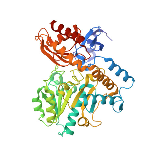
7TED, 7TEV, 7TFP - PubMed Abstract:
Human ornithine aminotransferase (hOAT) is a pyridoxal 5'-phosphate (PLP)-dependent enzyme that contains a similar active site to that of γ-aminobutyric acid aminotransferase (GABA-AT). Recently, pharmacological inhibition of hOAT was recognized as a potential therapeutic approach for hepatocellular carcinoma. In this work, we first studied the inactivation mechanisms of hOAT by two well-known GABA-AT inactivators ( CPP-115 and OV329 ). Inspired by the inactivation mechanistic difference between these two aminotransferases, a series of analogues were designed and synthesized, leading to the discovery of analogue 10b as a highly selective and potent hOAT inhibitor. Intact protein mass spectrometry, protein crystallography, and dialysis experiments indicated that 10b was converted to an irreversible tight-binding adduct ( 34 ) in the active site of hOAT, as was the unsaturated analogue ( 11 ). The comparison of kinetic studies between 10b and 11 suggested that the active intermediate ( 17b ) was only generated in hOAT and not in GABA-AT. Molecular docking studies and p K a computational calculations highlighted the importance of chirality and the endocyclic double bond for inhibitory activity. The turnover mechanism of 10b was supported by mass spectrometric analysis of dissociable products and fluoride ion release experiments. Notably, the stopped-flow experiments were highly consistent with the proposed mechanism, suggesting a relatively slow hydrolysis rate for hOAT. The novel second-deprotonation mechanism of 10b contributes to its high potency and significantly enhanced selectivity for hOAT inhibition.
- Department of Chemistry, Chemistry of Life Processes Institute, and Center for Developmental Therapeutics, Northwestern University, Evanston, Illinois 60208, United States.
Organizational Affiliation: