7TDL | pdb_00007tdl

M379A mutant tyrosine phenol-lyase complexed with 3-bromo-DL-phenylalanine

  • Classification: LYASE
  • Organism(s): Citrobacter freundii
  • Expression System: Escherichia coli
  • Mutation(s): Yes 

  • Deposited: 2022-01-01 Released: 2022-06-01 
  • Deposition Author(s): Phillips, R.S.
  • Funding Organization(s): National Institutes of Health/National Institute of General Medical Sciences (NIH/NIGMS)

Experimental Data Snapshot

  • Method: X-RAY DIFFRACTION
  • Resolution: 1.60 Å
  • R-Value Free: 
    0.221 (Depositor), 0.222 (DCC) 
  • R-Value Work: 
    0.177 (Depositor), 0.178 (DCC) 
  • R-Value Observed: 
    0.178 (Depositor) 

Starting Model: experimental
View more details

wwPDB Validation 3D Report Full Report

Validation slider image for 7TDL

Ligand Structure Quality Assessment 


This is version 1.3 of the entry. See complete history

Literature

M379A Mutant Tyrosine Phenol-lyase from Citrobacter freundii Has Altered Conformational Dynamics.

Phillips, R.S.Jones, B.Nash, S.

(2022) Chembiochem 23: e202200028-e202200028

  • DOI: https://doi.org/10.1002/cbic.202200028
  • Primary Citation Related Structures: 
    7TCS, 7TDL

  • PubMed Abstract: 

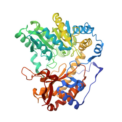
    The M379A mutant of Citrobacter freundii tyrosine phenol-lyase (TPL) has been prepared. M379A TPL is a robust catalyst to prepare a number of tyrosines substituted at the 3-position with bulky groups that cannot be made with wild type TPL. The three dimensional structures of M379A TPL complexed with L-methionine and 3-bromo-DL-phenylalanine have been determined by X-ray crystallography. Methionine is bound as a quinonoid complex in a closed active site in 3 of 4 chains of homotetrameric M379A TPL. M379A TPL reacts with L-methionine about 8-fold slower than wild type TPL. The temperature dependence shows that the slower reaction is due to less positive activation entropy. The structure of the M379A TPL complex of 3-bromo-DL-phenylalanine has a quinonoid complex in two subunits, with an open active site conformation. The effects of the M379A mutation on TPL suggest that the mutant enzyme has altered the conformational dynamics of the active site.


  • Organizational Affiliation
    • Department of Chemistry, University of Georgia, Athens, Georgia, 30602, USA.

Macromolecule Content 

  • Total Structure Weight: 208.06 kDa 
  • Atom Count: 16,411 
  • Modeled Residue Count: 1,824 
  • Deposited Residue Count: 1,824 
  • Unique protein chains: 1

Macromolecules

Find similar proteins by:|  3D Structure
Entity ID: 1
MoleculeChains  Sequence LengthOrganismDetailsImage
Tyrosine phenol-lyase
A, B, C, D
456Citrobacter freundiiMutation(s): 1 
Gene Names: tpl
EC: 4.1.99.2
UniProt
Find proteins for P31013 (Citrobacter freundii)
Explore P31013 
Go to UniProtKB:  P31013
Entity Groups
Sequence Clusters30% Identity50% Identity70% Identity90% Identity95% Identity100% Identity
UniProt GroupP31013
Sequence Annotations
Expand
Reference Sequence

Small Molecules

Ligands 5 Unique
IDChains Name / Formula / InChI Key2D Diagram3D Interactions
OY3
(Subject of Investigation/LOI)

Query on OY3


Download:Ideal Coordinates CCD File
G [auth A],
J [auth B]
(4Z)-4-({[(1E)-2-(3-bromophenyl)-1-carboxyethylidene]azaniumyl}methylidene)-2-methyl-5-[(phosphonooxy)methyl]-1,4-dihydropyridin-3-olate
C17 H18 Br N2 O7 P
QODYJTOPURJJAH-MTTBYZIPSA-N
PLP

Query on PLP


Download:Ideal Coordinates CCD File
K [auth B],
P [auth C],
S [auth D]
PYRIDOXAL-5'-PHOSPHATE
C8 H10 N O6 P
NGVDGCNFYWLIFO-UHFFFAOYSA-N
DMS

Query on DMS


Download:Ideal Coordinates CCD File
H [auth A],
L [auth C],
O [auth C],
R [auth D]
DIMETHYL SULFOXIDE
C2 H6 O S
IAZDPXIOMUYVGZ-UHFFFAOYSA-N
K

Query on K


Download:Ideal Coordinates CCD File
E [auth A],
F [auth A],
M [auth C],
N [auth C]
POTASSIUM ION
K
NPYPAHLBTDXSSS-UHFFFAOYSA-N
CL

Query on CL


Download:Ideal Coordinates CCD File
I [auth A],
Q [auth C],
T [auth D]
CHLORIDE ION
Cl
VEXZGXHMUGYJMC-UHFFFAOYSA-M

Experimental Data & Validation

Experimental Data

  • Method: X-RAY DIFFRACTION
  • Resolution: 1.60 Å
  • R-Value Free:  0.221 (Depositor), 0.222 (DCC) 
  • R-Value Work:  0.177 (Depositor), 0.178 (DCC) 
  • R-Value Observed: 0.178 (Depositor) 
Space Group: P 1
Unit Cell:
Length ( Å )Angle ( ˚ )
a = 61.25α = 113.16
b = 82.87β = 96.54
c = 94.33γ = 102.21
Software Package:
Software NamePurpose
PHENIXrefinement
autoPROCdata processing
XDSdata reduction
Aimlessdata scaling
PHASERphasing

Structure Validation

View Full Validation Report


Ligand Structure Quality Assessment 


Entry History 

& Funding Information

Deposition Data


Funding OrganizationLocationGrant Number
National Institutes of Health/National Institute of General Medical Sciences (NIH/NIGMS)United StatesR01-GM137008

Revision History  (Full details and data files)

  • Version 1.0: 2022-06-01
    Type: Initial release
  • Version 1.1: 2022-07-20
    Changes: Database references
  • Version 1.2: 2022-08-10
    Changes: Database references
  • Version 1.3: 2023-10-18
    Changes: Data collection, Refinement description