7TBH

cryo-EM structure of MBP-KIX-apoferritin complex with peptide 7


Experimental Data Snapshot

  • Method: ELECTRON MICROSCOPY
  • Resolution: 2.30 Å
  • Aggregation State: PARTICLE 
  • Reconstruction Method: SINGLE PARTICLE 

wwPDB Validation   3D Report Full Report


This is version 1.0 of the entry. See complete history


Literature

Cryo-EM, Protein Engineering, and Simulation Enable the Development of Peptide Therapeutics against Acute Myeloid Leukemia.

Zhang, K.Horikoshi, N.Li, S.Powers, A.S.Hameedi, M.A.Pintilie, G.D.Chae, H.D.Khan, Y.A.Suomivuori, C.M.Dror, R.O.Sakamoto, K.M.Chiu, W.Wakatsuki, S.

(2022) ACS Cent Sci 8: 214-222

  • DOI: https://doi.org/10.1021/acscentsci.1c01090
  • Primary Citation of Related Structures:  
    7TB3, 7TBH

  • PubMed Abstract: 

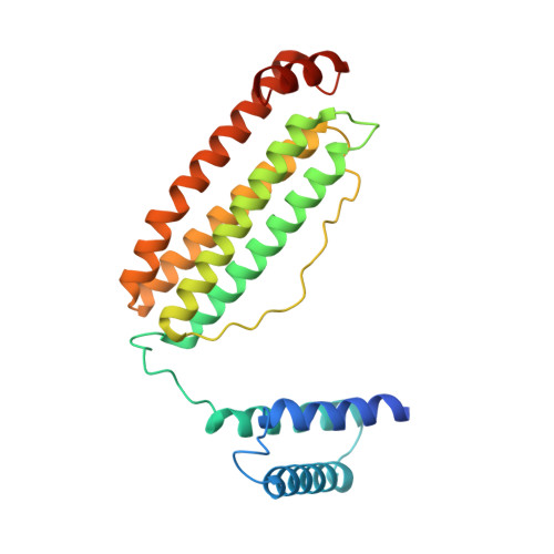
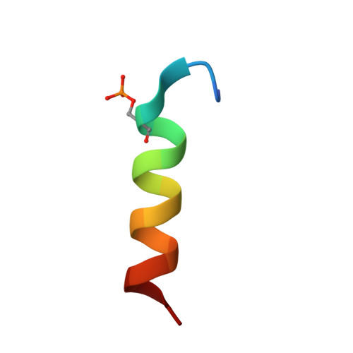
    Cryogenic electron microscopy (cryo-EM) has emerged as a viable structural tool for molecular therapeutics development against human diseases. However, it remains a challenge to determine structures of proteins that are flexible and smaller than 30 kDa. The 11 kDa KIX domain of CREB-binding protein (CBP), a potential therapeutic target for acute myeloid leukemia and other cancers, is a protein which has defied structure-based inhibitor design. Here, we develop an experimental approach to overcome the size limitation by engineering a protein double-shell to sandwich the KIX domain between apoferritin as the inner shell and maltose-binding protein as the outer shell. To assist homogeneous orientations of the target, disulfide bonds are introduced at the target-apoferritin interface, resulting in a cryo-EM structure at 2.6 Å resolution. We used molecular dynamics simulations to design peptides that block the interaction of the KIX domain of CBP with the intrinsically disordered pKID domain of CREB. The double-shell design allows for fluorescence polarization assays confirming the binding between the KIX domain in the double-shell and these interacting peptides. Further cryo-EM analysis reveals a helix-helix interaction between a single KIX helix and the best peptide, providing a possible strategy for developments of next-generation inhibitors.


  • Organizational Affiliation

    MOE Key Laboratory for Cellular Dynamics and Division of Life Sciences and Medicine, University of Science and Technology of China, Hefei 230027, China.


Macromolecules
Find similar proteins by:  (by identity cutoff)  |  3D Structure
Entity ID: 1
MoleculeChains Sequence LengthOrganismDetailsImage
Isoform 2 of CREB-binding protein,Ferritin heavy chain, N-terminally processed
A, B, C, D, E
A, B, C, D, E, F, G, H, I, J, K, L, M, N, O, P, Q, R, S, T, U, V, W, X
260Homo sapiensMutation(s): 0 
Gene Names: CREBBPCBPFth1Fth
EC: 2.3.1.48 (PDB Primary Data), 2.3.1 (PDB Primary Data)
UniProt & NIH Common Fund Data Resources
Find proteins for P09528 (Mus musculus)
Explore P09528 
Go to UniProtKB:  P09528
IMPC:  MGI:95588
Find proteins for Q92793 (Homo sapiens)
Explore Q92793 
Go to UniProtKB:  Q92793
PHAROS:  Q92793
GTEx:  ENSG00000005339 
Entity Groups  
Sequence Clusters30% Identity50% Identity70% Identity90% Identity95% Identity100% Identity
UniProt GroupsQ92793P09528
Sequence Annotations
Expand
  • Reference Sequence

Find similar proteins by:  Sequence   |   3D Structure  

Entity ID: 2
MoleculeChains Sequence LengthOrganismDetailsImage
LEU-SER-ARG-ARG-PRO-SEP-TYR-ARG-LYS-ILE-LEU-ASN-ASP-LEU-SER-SER-ASP-ALA-PRO19Homo sapiensMutation(s): 0 
UniProt & NIH Common Fund Data Resources
Find proteins for P16220 (Homo sapiens)
Explore P16220 
Go to UniProtKB:  P16220
PHAROS:  P16220
GTEx:  ENSG00000118260 
Entity Groups  
Sequence Clusters30% Identity50% Identity70% Identity90% Identity95% Identity100% Identity
UniProt GroupP16220
Sequence Annotations
Expand
  • Reference Sequence
Small Molecules
Modified Residues  1 Unique
IDChains TypeFormula2D DiagramParent
SEP
Query on SEP
AA [auth c]
BA [auth d]
CA [auth e]
DA [auth f]
EA [auth g]
AA [auth c],
BA [auth d],
CA [auth e],
DA [auth f],
EA [auth g],
FA [auth h],
GA [auth i],
HA [auth j],
IA [auth k],
JA [auth l],
KA [auth m],
LA [auth n],
MA [auth o],
NA [auth p],
OA [auth q],
PA [auth r],
QA [auth s],
RA [auth t],
SA [auth u],
TA [auth v],
UA [auth w],
VA [auth x],
Y [auth a],
Z [auth b]
L-PEPTIDE LINKINGC3 H8 N O6 PSER
Experimental Data & Validation

Experimental Data

  • Method: ELECTRON MICROSCOPY
  • Resolution: 2.30 Å
  • Aggregation State: PARTICLE 
  • Reconstruction Method: SINGLE PARTICLE 
EM Software:
TaskSoftware PackageVersion
MODEL REFINEMENTPHENIX
RECONSTRUCTIONcryoSPARC2

Structure Validation

View Full Validation Report


Entry History & Funding Information

Deposition Data


Funding OrganizationLocationGrant Number
National Institutes of Health/National Institute of General Medical Sciences (NIH/NIGMS)United StatesR01GM079429
National Institutes of Health/National Institute of General Medical Sciences (NIH/NIGMS)United StatesP41GM103832
National Institutes of Health/National Institute of General Medical Sciences (NIH/NIGMS)United StatesS10OD021600

Revision History  (Full details and data files)

  • Version 1.0: 2022-03-16
    Type: Initial release