Insight into the mechanism of H + -coupled nucleobase transport.
Weng, J., Zhou, X., Wiriyasermkul, P., Ren, Z., Chen, K., Gil-Iturbe, E., Zhou, M., Quick, M.(2023) Proc Natl Acad Sci U S A 120: e2302799120-e2302799120
- PubMed: 37549264 Search on PubMedSearch on PubMed Central
- DOI: https://doi.org/10.1073/pnas.2302799120
- Primary Citation Related Structures:
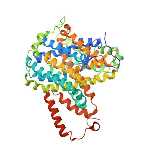
7TAK - PubMed Abstract:
Members of the nucleobase/ascorbic acid transporter (NAT) gene family are found in all kingdoms of life. In mammals, the concentrative uptake of ascorbic acid (vitamin C) by members of the NAT family is driven by the Na + gradient, while the uptake of nucleobases in bacteria is powered by the H + gradient. Here, we report the structure and function of PurT Cp , a NAT family member from Colwellia psychrerythraea . The structure of PurT Cp was determined to 2.80 Å resolution by X-ray crystallography. PurT Cp forms a homodimer, and each protomer has 14 transmembrane segments folded into a transport domain (core domain) and a scaffold domain (gate domain). A purine base is present in the structure and defines the location of the substrate binding site. Functional studies reveal that PurT Cp transports purines but not pyrimidines and that purine binding and transport is dependent on the pH. Mutation of a conserved aspartate residue close to the substrate binding site reveals the critical role of this residue in H + -dependent transport of purines. Comparison of the PurT Cp structure with transporters of the same structural fold suggests that rigid-body motions of the substrate-binding domain are central for substrate translocation across the membrane.
- Department of Physiology and Cellular Biophysics, Columbia University Irving Medical Center, New York, NY 10032.
Organizational Affiliation: