7T9L | pdb_00007t9l

Cryo-EM structure of SARS-CoV-2 Omicron spike protein in complex with human ACE2 (focused refinement of RBD and ACE2)


Experimental Data Snapshot

  • Method: ELECTRON MICROSCOPY
  • Resolution: 2.66 Å
  • Aggregation State: PARTICLE 
  • Reconstruction Method: SINGLE PARTICLE 

wwPDB Validation 3D Report Full Report

Validation slider image for 7T9L

This is version 1.2 of the entry. See complete history

Literature

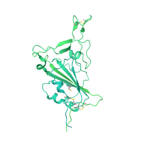
SARS-CoV-2 Omicron variant: Antibody evasion and cryo-EM structure of spike protein-ACE2 complex.

Mannar, D.Saville, J.W.Zhu, X.Srivastava, S.S.Berezuk, A.M.Tuttle, K.S.Marquez, A.C.Sekirov, I.Subramaniam, S.

(2022) Science 375: 760-764

  • DOI: https://doi.org/10.1126/science.abn7760
  • Primary Citation Related Structures: 
    7T9J, 7T9K, 7T9L

  • PubMed Abstract: 

    The newly reported Omicron variant is poised to replace Delta as the most prevalent SARS-CoV-2 variant across the world. Cryo-EM structural analysis of the Omicron variant spike protein in complex with human ACE2 reveals new salt bridges and hydrogen bonds formed by mutated residues R493, S496 and R498 in the RBD with ACE2. These interactions appear to compensate for other Omicron mutations such as K417N known to reduce ACE2 binding affinity, resulting in similar biochemical ACE2 binding affinities for Delta and Omicron variants. Neutralization assays show that pseudoviruses displaying the Omicron spike protein exhibit increased antibody evasion. The increase in antibody evasion, together with retention of strong interactions at the ACE2 interface, thus represent important molecular features that likely contribute to the rapid spread of the Omicron variant.


  • Organizational Affiliation
    • Department of Biochemistry and Molecular Biology, University of British Columbia, Vancouver, BC, Canada.

Macromolecule Content 

  • Total Structure Weight: 214.52 kDa 
  • Atom Count: 6,567 
  • Modeled Residue Count: 796 
  • Deposited Residue Count: 1,891 
  • Unique protein chains: 2

Macromolecules

Find similar proteins by:|  3D Structure
Entity ID: 1
MoleculeChains  Sequence LengthOrganismDetailsImage
Spike glycoprotein1,285Severe acute respiratory syndrome coronavirus 2Mutation(s): 10 
Gene Names: S2
UniProt
Find proteins for P0DTC2 (Severe acute respiratory syndrome coronavirus 2)
Explore P0DTC2 
Go to UniProtKB:  P0DTC2
Entity Groups
Sequence Clusters30% Identity50% Identity70% Identity90% Identity95% Identity100% Identity
UniProt GroupP0DTC2
Glycosylation
Glycosylation Sites: 1Go to GlyGen: P0DTC2-1
Sequence Annotations
Expand
Reference Sequence
Find similar proteins by:|  3D Structure
Entity ID: 2
MoleculeChains  Sequence LengthOrganismDetailsImage
Processed angiotensin-converting enzyme 2B [auth D]606Homo sapiensMutation(s): 0 
Gene Names: ACE2UNQ868/PRO1885
EC: 3.4.17 (UniProt), 3.4.17.23 (UniProt)
UniProt & NIH Common Fund Data Resources
Find proteins for Q9BYF1 (Homo sapiens)
Explore Q9BYF1 
Go to UniProtKB:  Q9BYF1
PHAROS:  Q9BYF1
GTEx:  ENSG00000130234 
Entity Groups
Sequence Clusters30% Identity50% Identity70% Identity90% Identity95% Identity100% Identity
UniProt GroupQ9BYF1
Glycosylation
Glycosylation Sites: 6Go to GlyGen: Q9BYF1-1
Sequence Annotations
Expand
Reference Sequence

Experimental Data & Validation

Experimental Data

  • Method: ELECTRON MICROSCOPY
  • Resolution: 2.66 Å
  • Aggregation State: PARTICLE 
  • Reconstruction Method: SINGLE PARTICLE 

Structure Validation

View Full Validation Report


Entry History 

& Funding Information

Deposition Data


Funding OrganizationLocationGrant Number
Canada Excellence Research Chair AwardPrecision Cancer Drug Design
Other privateCOVID-19 research

Revision History  (Full details and data files)

  • Version 1.0: 2021-12-29
    Type: Initial release
  • Version 1.1: 2022-07-13
    Changes: Database references
  • Version 1.2: 2024-11-13
    Changes: Data collection, Structure summary