7SZH

Structure of the Rieske Non-heme Iron Oxygenase SxtT with beta-saxitoxinol Bound


Experimental Data Snapshot

  • Method: X-RAY DIFFRACTION
  • Resolution: 1.79 Å
  • R-Value Free: 0.199 
  • R-Value Work: 0.171 
  • R-Value Observed: 0.172 

wwPDB Validation   3D Report Full Report


Ligand Structure Quality Assessment 


This is version 1.2 of the entry. See complete history


Literature

Design principles for site-selective hydroxylation by a Rieske oxygenase.

Liu, J.Tian, J.Perry, C.Lukowski, A.L.Doukov, T.I.Narayan, A.R.H.Bridwell-Rabb, J.

(2022) Nat Commun 13: 255-255

  • DOI: https://doi.org/10.1038/s41467-021-27822-3
  • Primary Citation of Related Structures:  
    7SZE, 7SZF, 7SZG, 7SZH

  • PubMed Abstract: 

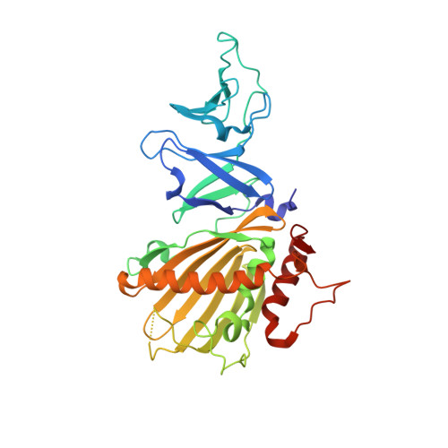
    Rieske oxygenases exploit the reactivity of iron to perform chemically challenging C-H bond functionalization reactions. Thus far, only a handful of Rieske oxygenases have been structurally characterized and remarkably little information exists regarding how these enzymes use a common architecture and set of metallocenters to facilitate a diverse range of reactions. Herein, we detail how two Rieske oxygenases SxtT and GxtA use different protein regions to influence the site-selectivity of their catalyzed monohydroxylation reactions. We present high resolution crystal structures of SxtT and GxtA with the native β-saxitoxinol and saxitoxin substrates bound in addition to a Xenon-pressurized structure of GxtA that reveals the location of a substrate access tunnel to the active site. Ultimately, this structural information allowed for the identification of six residues distributed between three regions of SxtT that together control the selectivity of the C-H hydroxylation event. Substitution of these residues produces a SxtT variant that is fully adapted to exhibit the non-native site-selectivity and substrate scope of GxtA. Importantly, we also found that these selectivity regions are conserved in other structurally characterized Rieske oxygenases, providing a framework for predictively repurposing and manipulating Rieske oxygenases as biocatalysts.


  • Organizational Affiliation

    Department of Chemistry, University of Michigan, Ann Arbor, MI, 48109, USA.


Macromolecules
Find similar proteins by:  (by identity cutoff)  |  3D Structure
Entity ID: 1
MoleculeChains Sequence LengthOrganismDetailsImage
SxtT
A, B, C
334Microseira wolleiMutation(s): 0 
Gene Names: sxtT
UniProt
Find proteins for C3RVQ0 (Microseira wollei)
Explore C3RVQ0 
Go to UniProtKB:  C3RVQ0
Entity Groups  
Sequence Clusters30% Identity50% Identity70% Identity90% Identity95% Identity100% Identity
UniProt GroupC3RVQ0
Sequence Annotations
Expand
  • Reference Sequence
Small Molecules
Ligands 6 Unique
IDChains Name / Formula / InChI Key2D Diagram3D Interactions
D82 (Subject of Investigation/LOI)
Query on D82

Download Ideal Coordinates CCD File 
I [auth A],
IA [auth C],
Y [auth B]
beta-Saxitoxinol
C10 H17 N7 O3
NILHUXIFTLLDPJ-AVGUDYQDSA-N
FES (Subject of Investigation/LOI)
Query on FES

Download Ideal Coordinates CCD File 
D [auth A],
DA [auth C],
O [auth B]
FE2/S2 (INORGANIC) CLUSTER
Fe2 S2
NIXDOXVAJZFRNF-UHFFFAOYSA-N
SO4 (Subject of Investigation/LOI)
Query on SO4

Download Ideal Coordinates CCD File 
AA [auth B]
BA [auth B]
CA [auth B]
K [auth A]
OA [auth C]
AA [auth B],
BA [auth B],
CA [auth B],
K [auth A],
OA [auth C],
PA [auth C]
SULFATE ION
O4 S
QAOWNCQODCNURD-UHFFFAOYSA-L
GOL (Subject of Investigation/LOI)
Query on GOL

Download Ideal Coordinates CCD File 
E [auth A]
EA [auth C]
F [auth A]
FA [auth C]
G [auth A]
E [auth A],
EA [auth C],
F [auth A],
FA [auth C],
G [auth A],
GA [auth C],
H [auth A],
HA [auth C],
JA [auth C],
KA [auth C],
LA [auth C],
MA [auth C],
N [auth B],
P [auth B],
Q [auth B],
R [auth B],
S [auth B],
T [auth B],
U [auth B],
V [auth B],
W [auth B],
X [auth B]
GLYCEROL
C3 H8 O3
PEDCQBHIVMGVHV-UHFFFAOYSA-N
FE (Subject of Investigation/LOI)
Query on FE

Download Ideal Coordinates CCD File 
J [auth A],
NA [auth C],
Z [auth B]
FE (III) ION
Fe
VTLYFUHAOXGGBS-UHFFFAOYSA-N
CL (Subject of Investigation/LOI)
Query on CL

Download Ideal Coordinates CCD File 
L [auth A],
M [auth A]
CHLORIDE ION
Cl
VEXZGXHMUGYJMC-UHFFFAOYSA-M
Experimental Data & Validation

Experimental Data

  • Method: X-RAY DIFFRACTION
  • Resolution: 1.79 Å
  • R-Value Free: 0.199 
  • R-Value Work: 0.171 
  • R-Value Observed: 0.172 
  • Space Group: C 2 2 2
Unit Cell:
Length ( Å )Angle ( ˚ )
a = 153.282α = 90
b = 158.47β = 90
c = 115.839γ = 90
Software Package:
Software NamePurpose
PHENIXrefinement
XDSdata reduction
XDSdata scaling
PHENIXphasing

Structure Validation

View Full Validation Report


Ligand Structure Quality Assessment 


Entry History & Funding Information

Deposition Data


Funding OrganizationLocationGrant Number
National Institutes of Health/National Institute of General Medical Sciences (NIH/NIGMS)United StatesR35 GM138271

Revision History  (Full details and data files)

  • Version 1.0: 2022-01-19
    Type: Initial release
  • Version 1.1: 2022-01-26
    Changes: Database references
  • Version 1.2: 2023-10-18
    Changes: Data collection, Refinement description